Copyright
©The Author(s) 2024.
World J Gastroenterol. Apr 14, 2024; 30(14): 1949-1957
Published online Apr 14, 2024. doi: 10.3748/wjg.v30.i14.1949
Published online Apr 14, 2024. doi: 10.3748/wjg.v30.i14.1949
Figure 1 A typical histologic finding of chronic hepatitis C.
A dense, follicle-like accumulation of lymphocytes is seen in the portal area. Hematoxylin-eosin, original magnification 200 ×.
Figure 2 The number of liver biopsies conducted at Takatsuki General Hospital starting from 2011.
In 2014, oral direct-acting antiviral agents were introduced, resulting in a steady decline of liver biopsies.
Figure 3 Biopsy from 2 patients.
A and B: A liver biopsy from a 78-year-old male positive for antinuclear antibody. Hematoxylin-eosin (HE), original magnifications 40 × (A); HE, original magnifications 400 × (B); C-E: A liver biopsy from a 62-year-old male patient with diabetes mellitus. HE, original magnifications 100 × (C), 200 ×(D) and 200 ×(E).
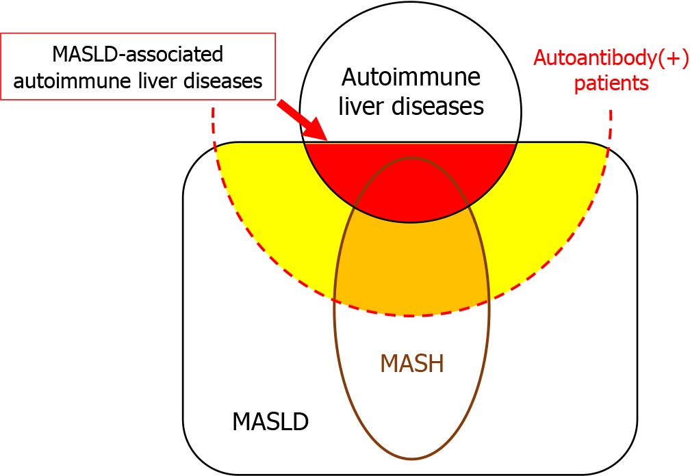
Figure 4 A schematic illustration indicating the relationships among metabolic-dysfunction associated steatotic liver disease, autoim
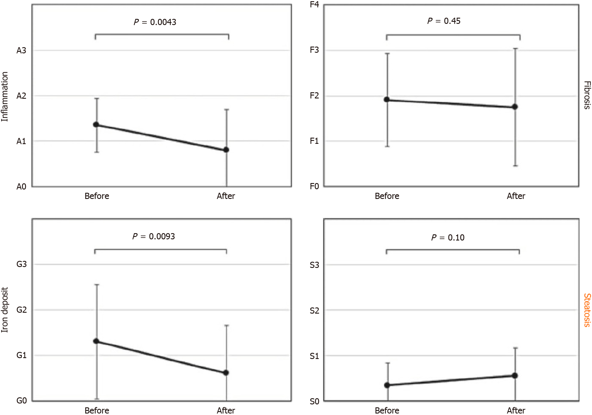
Figure 5 Findings of comparative biopsies before and after hepatitis C virus eradication with direct-acting antiviral agents.
While inflammation, fibrosis and iron deposition have improved, steatosis has not changed and appears to have worsened[8].
Figure 6 A typical case of hepatitis C virus-associated hepatocellular carcinoma.
A: Hematoxylin-eosin (HE), original magnifications 20 ×; B: HE, original magnifications 100 ×.
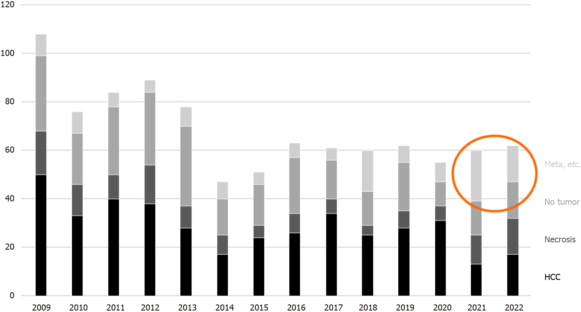
Figure 7 Trends in the number of tumor biopsies at Kansai Medical University Medical Center.
Meta: Metastatic tumor; HCC: Hepatocellular carcinoma.
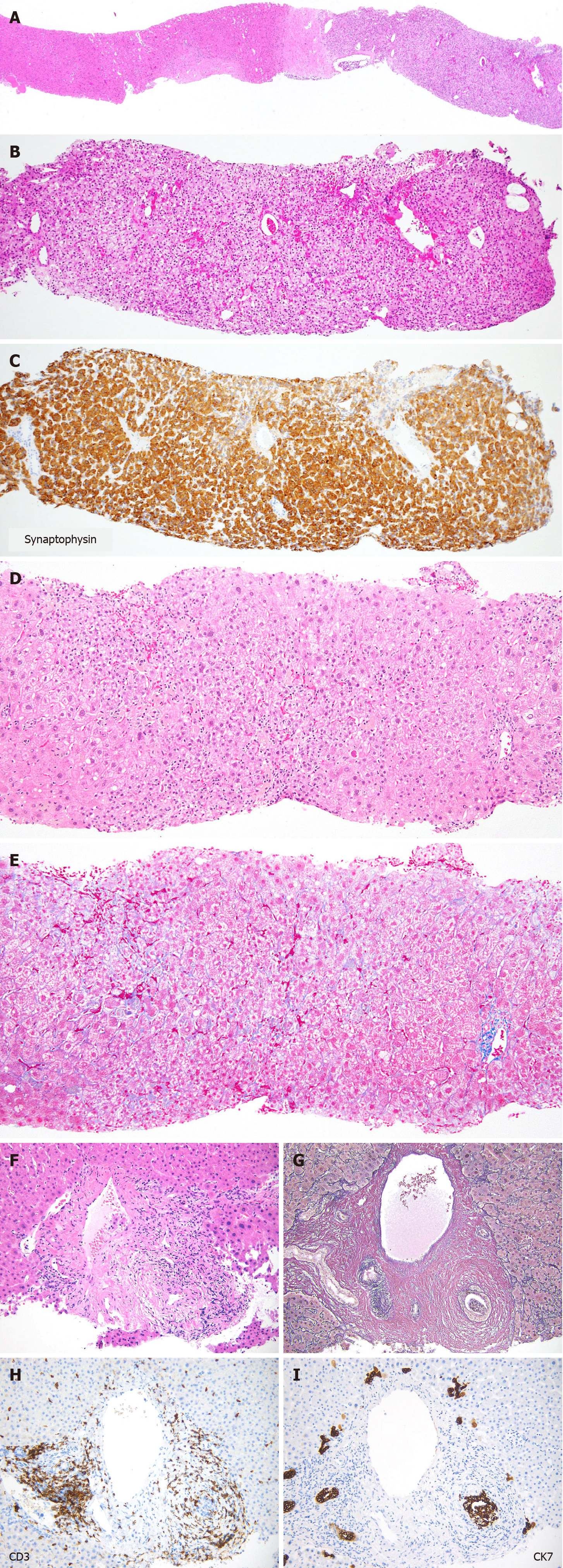
Figure 8 Liver biopsy from 3 patients.
A-C: A biopsy of hepatic tumor from a 60-year-old female patient previously diagnosed as hepatocellular carcinoma. Hematoxylin-eosin (HE), original magnifications 20 × (A), 100 × (B); Immunostaining for synaptoophysin, original magnification 100 × (C); D and E: A liver biopsy from a 72-year-old female patient diagnosed as drug-induced liver injury. HE, original magnification 100 × (D); Azan-Mallory stain, original magnification 100 × (E); F-I: A liver biopsy from a 76-year-old male patient with bile duct injury due to inhibitors-related adverse effects. HE, original magnification 100 × (F); Elastica van Gieson, original magnification 100 × (G); Immunostaining for CD3 and cytokeratin 7, original magnification 100 × (H, I). CK7: Cytokeratin 7.
- Citation: Ikura Y, Okubo T, Sakai Y. Liver biopsy in the post-hepatitis C virus era in Japan. World J Gastroenterol 2024; 30(14): 1949-1957
- URL: https://www.wjgnet.com/1007-9327/full/v30/i14/1949.htm
- DOI: https://dx.doi.org/10.3748/wjg.v30.i14.1949


