Copyright
©The Author(s) 2024.
World J Gastroenterol. Apr 14, 2024; 30(14): 1934-1940
Published online Apr 14, 2024. doi: 10.3748/wjg.v30.i14.1934
Published online Apr 14, 2024. doi: 10.3748/wjg.v30.i14.1934
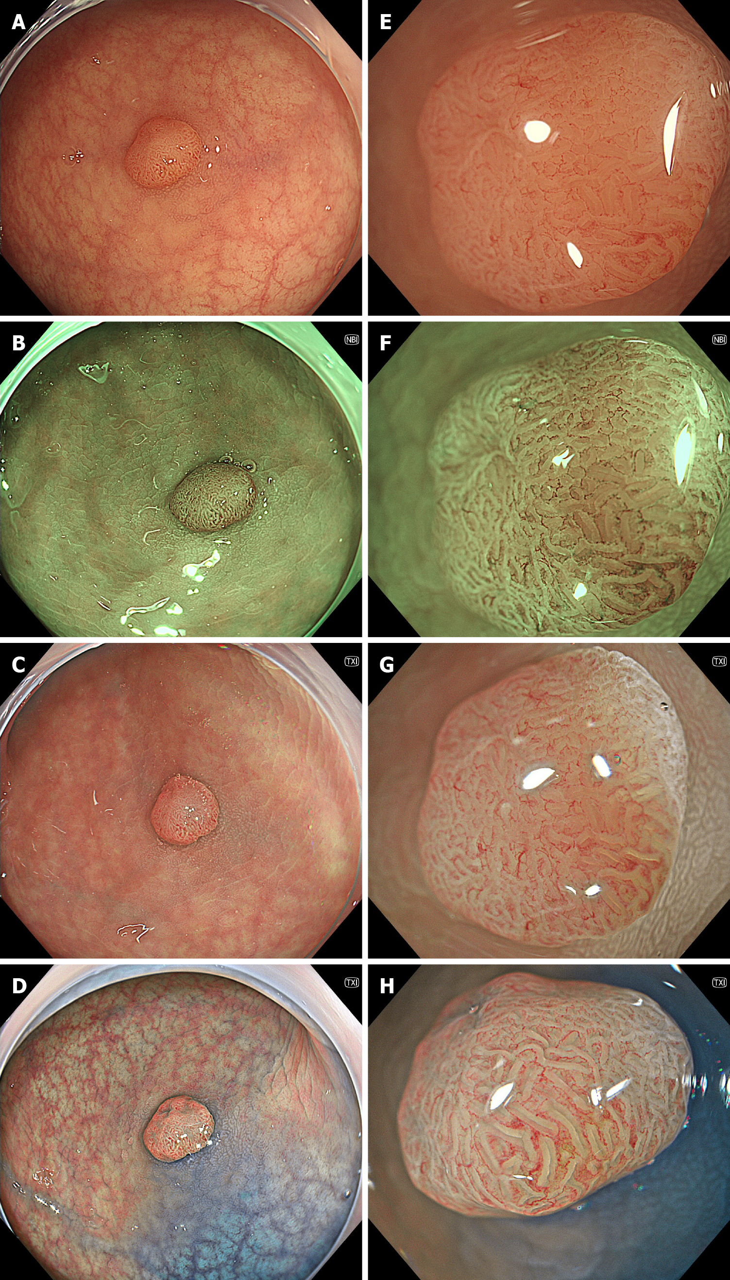
Figure 1 Representative endoscopic images of a colonic adenoma.
A-D: Conventional view; E-H: Magnifying view. A and E: White-light imaging; B and F: Narrow-band imaging; C and G: Texture and color enhancement imaging; D and H: Texture and color enhancement imaging with indigo carmine. An Olympus’ X1 system and a CF-XZ1200 scope were used.
- Citation: Toyoshima O, Nishizawa T, Hata K. Topic highlight on texture and color enhancement imaging in gastrointestinal diseases. World J Gastroenterol 2024; 30(14): 1934-1940
- URL: https://www.wjgnet.com/1007-9327/full/v30/i14/1934.htm
- DOI: https://dx.doi.org/10.3748/wjg.v30.i14.1934


