Copyright
©The Author(s) 1997.
World J Gastroenterol. Jun 15, 1997; 3(2): 87-89
Published online Jun 15, 1997. doi: 10.3748/wjg.v3.i2.87
Published online Jun 15, 1997. doi: 10.3748/wjg.v3.i2.87
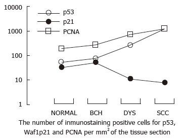
Figure 1 Changes in p53 and Waf1p21 expression in normal esophageal epithelia and epithelia with different degrees of lesions.
As the lesions progressed from BCH, DYS to SCC, the number of p53- and PCNA-positive cells significantly increased. In contrast, the number of Waf1p21-positive cells slightly increased from normal to BCH, but there was no further increase in DYS and in SCC. SCC: Squamous cell carcinoma; DYS: Dysplasia; BCH: Basal cell hyperplasia.
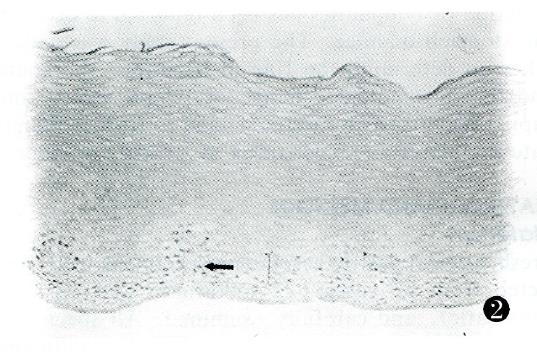
Figure 2 Waf1p21 immunostaining in biopsy samples of esophageal BCH.
Waf1p21-positive cells localized to the third and fourth cell epithelial layers (arrow). Scale bar, 2.4 μm. BCH: Basal cell hyperplasia.
- Citation: Wang LD, Yang WC, Zhou Q, Xing Y, Jia YY, Zhao X. Changes in p53 and Waf1p21 expression and cell proliferation in esophageal carcinogenesis. World J Gastroenterol 1997; 3(2): 87-89
- URL: https://www.wjgnet.com/1007-9327/full/v3/i2/87.htm
- DOI: https://dx.doi.org/10.3748/wjg.v3.i2.87


