Copyright
©The Author(s) 1997.
World J Gastroenterol. Jun 15, 1997; 3(2): 104-107
Published online Jun 15, 1997. doi: 10.3748/wjg.v3.i2.104
Published online Jun 15, 1997. doi: 10.3748/wjg.v3.i2.104
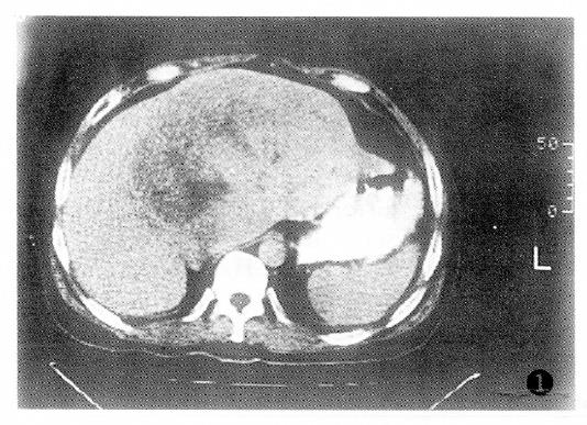
Figure 1 Before treatment, the computed tomography scan of the massive type of primary hepatic carcinoma showing local low density in the right and left hepatic lobar regions with 13 cm × 12 cm in size.
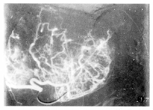
Figure 2 Before treatment, angiography of the patient showing a massive hypervascular tumor with its blood supply from the right and the left hepatic arteries.
HAI combined with HAE*Lp-BS treatment being performed. HAI: Hepatic arterial infusion chemotherapy; HAE: Hepatic arterial embolization.
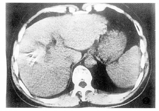
Figure 3 Eight months after treatment with HAI combined with HAE*Lp-BS, the computed tomography scan of the patient showed obvious tumor shrinking with 5 cm × 2.
5 cm in size and complete intratumor retention of lipiodol. HAI: Hepatic arterial infusion chemotherapy; HAE: Hepatic arterial embolization.
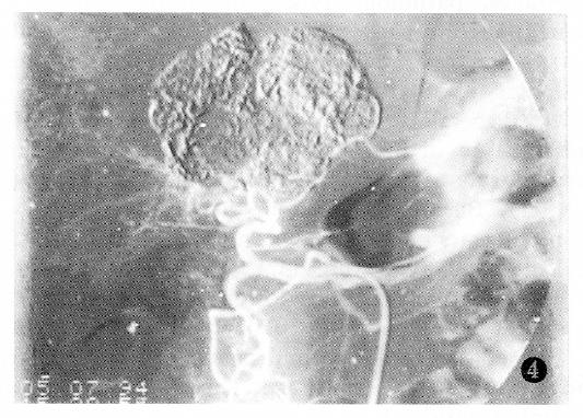
Figure 4 Eight months after treatment, angiography of the patient showed obvious tumor shrinking and complete retention of Lipiodol in the massive tumor and in the areas surrounding both small tumor nodules, without tumor vessels and collateral circulation.
There was no recanalization of the main trunk of the hepatic artery.
- Citation: Zheng CS, Feng GS, Zhou RM, Liang B, Liang HM, Zhen J, Yu JM, Liu H. Hepatic arterial infusion chemotherapy and embolization in the treatment of primary hepatic carcinoma. World J Gastroenterol 1997; 3(2): 104-107
- URL: https://www.wjgnet.com/1007-9327/full/v3/i2/104.htm
- DOI: https://dx.doi.org/10.3748/wjg.v3.i2.104


