Copyright
©The Author(s) 2023.
World J Gastroenterol. Feb 21, 2023; 29(7): 1173-1193
Published online Feb 21, 2023. doi: 10.3748/wjg.v29.i7.1173
Published online Feb 21, 2023. doi: 10.3748/wjg.v29.i7.1173
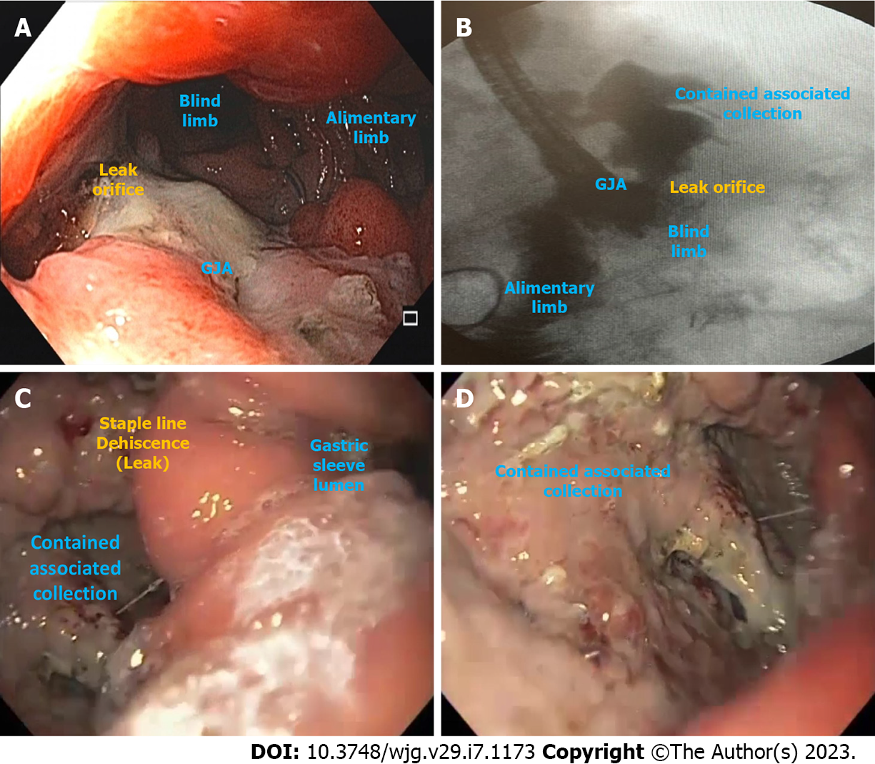
Figure 1 Post-bariatric surgical leaks illustrations.
A and B: Gastrojejunal anastomotic leak with an associated contained collection after Roux-en-Y gastric bypass; C and D: Leak with a contained associated collection due to staple line dehiscence after laparoscopic sleeve gastrectomy. GJA: Gastrojejunal anastomosis.
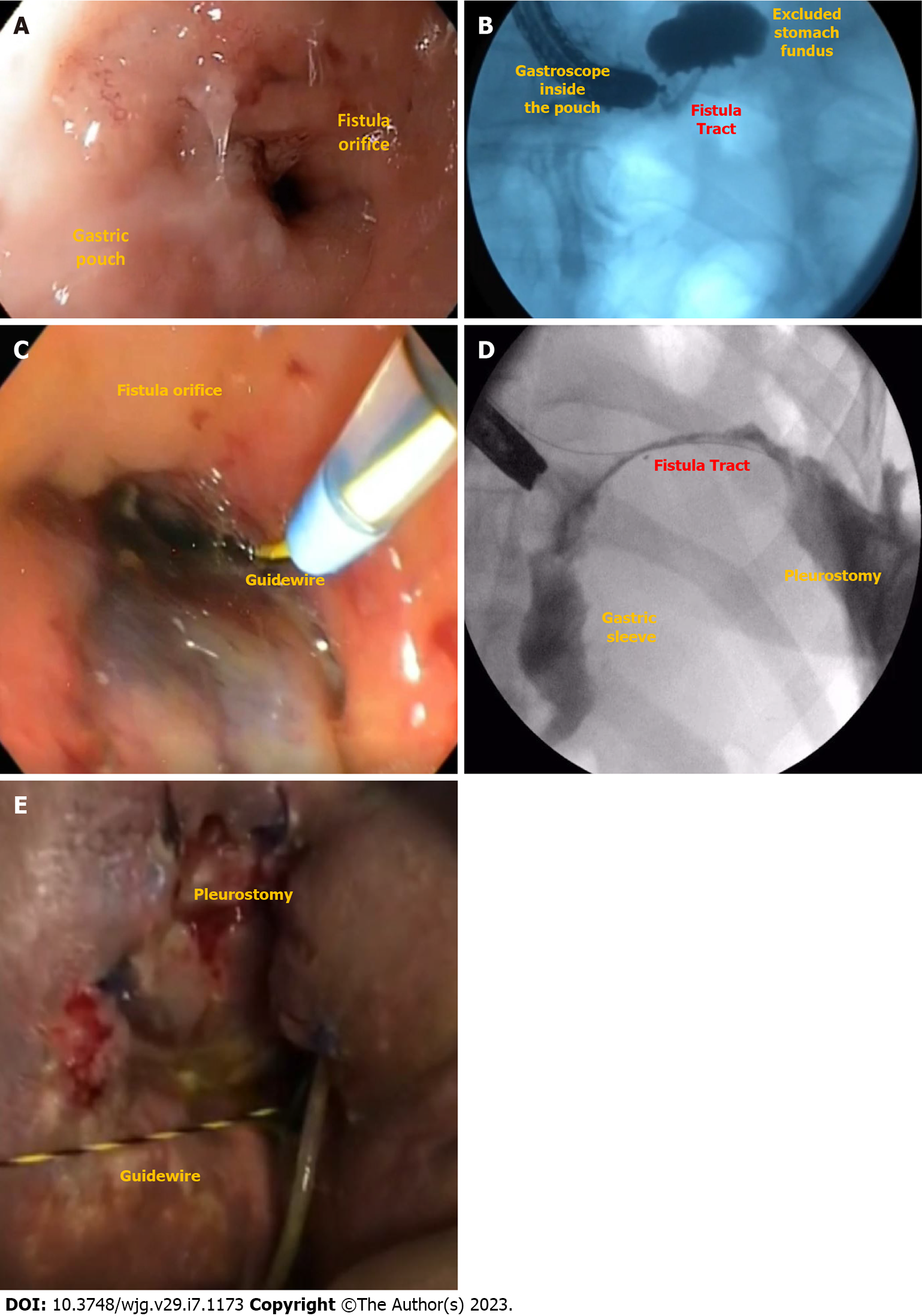
Figure 2 Post-bariatric surgical fistulas illustrations.
A and B: Gastrogastric fistula after Roux-en-Y gastric bypass; C, D, and E: Gastropleural fistula after laparoscopic sleeve gastrectomy.
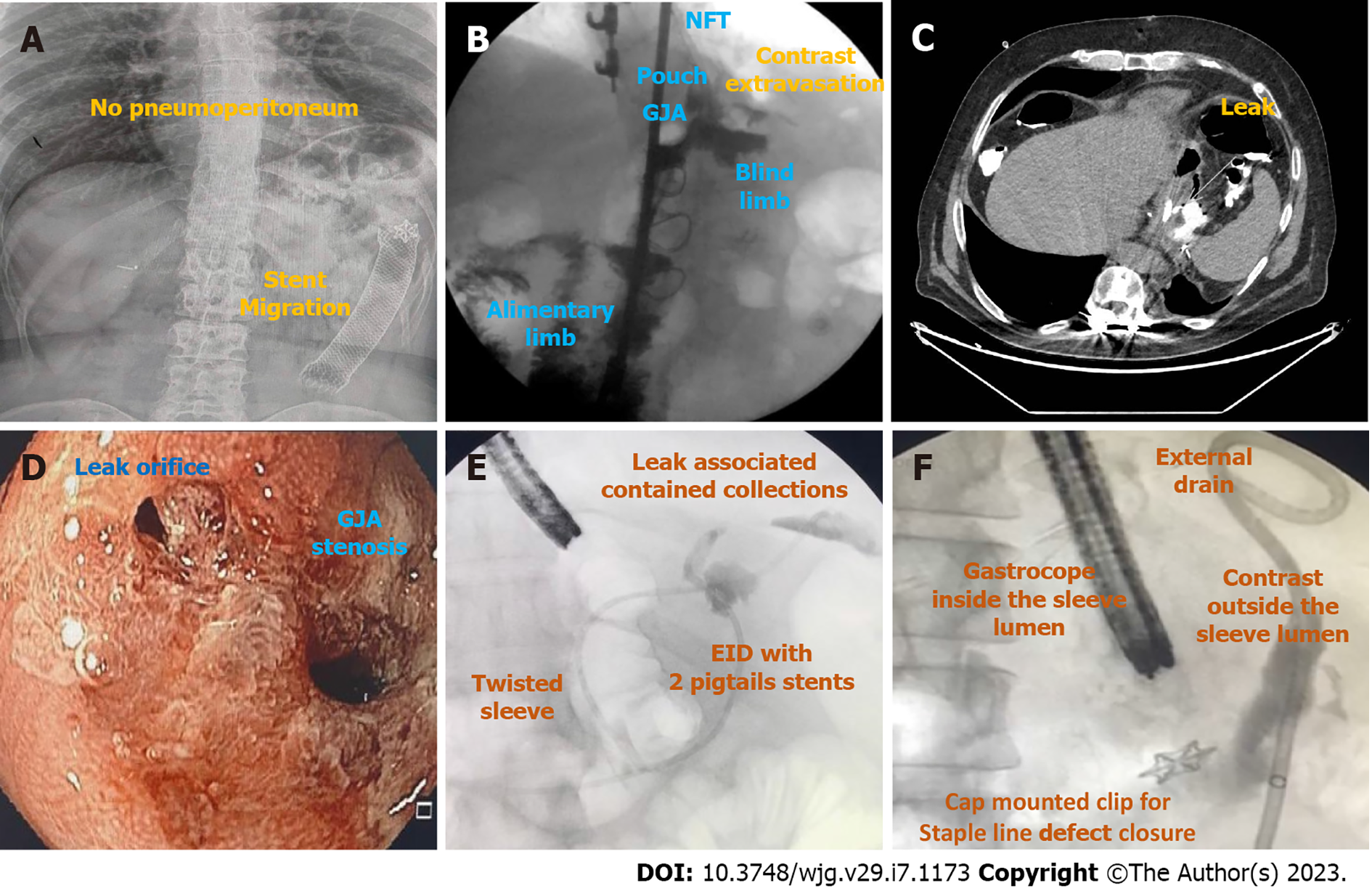
Figure 3 Imaging exams for diagnosis of leaks and fistulas after bariatric surgery.
A: Abdominal radiograph showing an esophageal fully covered metal stent fixed with a cap mounted clip at its proximal edge used for the treatment of a gastrojejunal anastomosis (GJA) leak after Roux-en-Y gastric bypass (RYGB) migrated to distal jejunum with no signs of pneuperitoneum; B: Upper gastrointestinal (GI) transit for suspicious leak after RYGB showing contrast extravasation; C: Computed tomography scan confirming the GJA leak suspected in the upper GI series (Figure 3B); D: Endoscopic visualization of a GJA stenosis associated with a leak; E: Fluoroscopy image during esophagogastroduodenoscopy (EGD) with injection of water soluble contrast through a catheter showing two leaks in the sleeve staple line with associated collections treated with endoscopic internal drainage with pigtail stents; F: Fluoroscopy image during EGD with injection of water soluble contrast through the external drain showing no extravasation for the intraluminal compartment, confirming the successful defect closure. GJA: Gastrojejunal anastomosis; EID: Endoscopic internal drainage; NFT: Nasoenteral feeding tube.
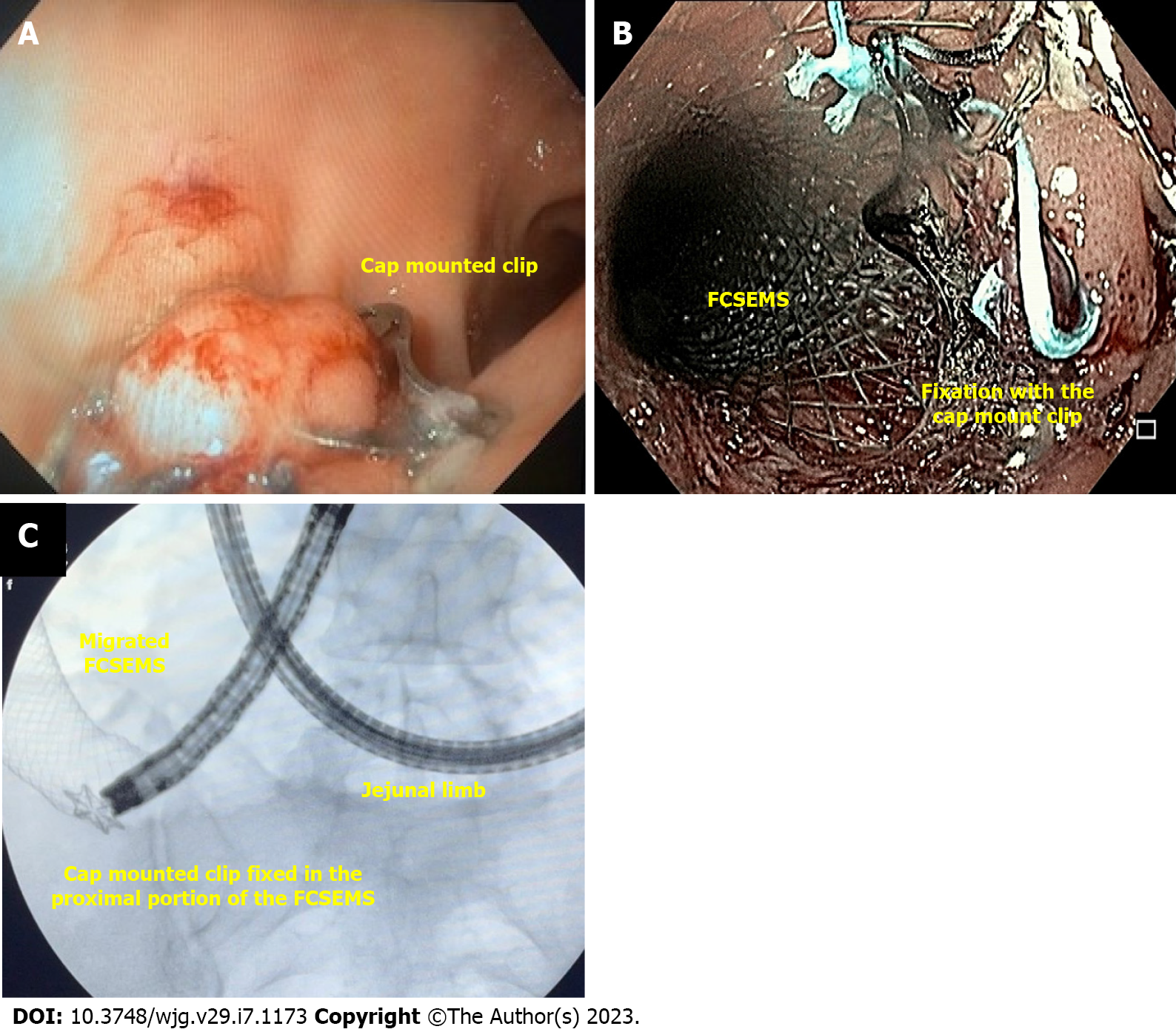
Figure 4 Cap-mounted clip.
A: Cap-mounted clip for an acute leak (orifice < 2 cm) in the staple line after laparoscopic sleeve gastrectomy in a stable patient with external drainage; B: Cap-mounted clip used for stent fixation; C: Enteroscopy to remove stent after migration due to cap mounted clip fixation failure. FCSEMS: Fully covered self-expandable metal stents.
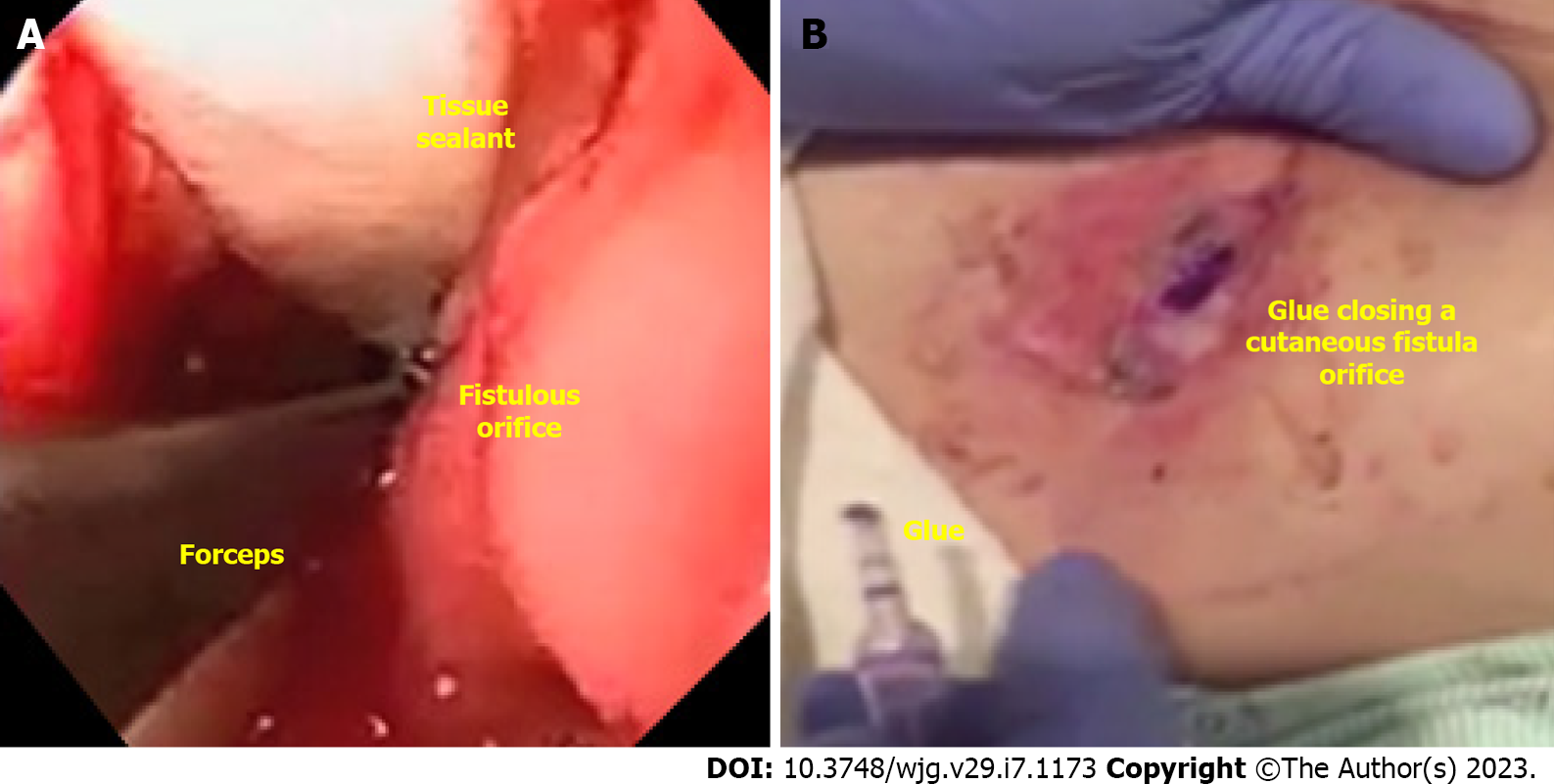
Figure 5 Tissue sealants/Glues.
A: Endoscopic placement of acellular matrix biomaterial in a late gastrocutaneous fistula after laparoscopic sleeve gastrectomy (LSG); B: Percutaneous application of glue (cyanoacrylate) in a chronic gastrocutaneous fistula tract after LSG.
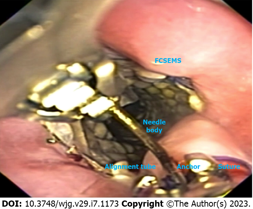
Figure 6 Endoscopic suturing for stent fixation placed in the distal esophagus of a patient with an early leak after Roux-en-Y gastric bypass.
FCSEMS: Fully covered self-expandable metal stents.
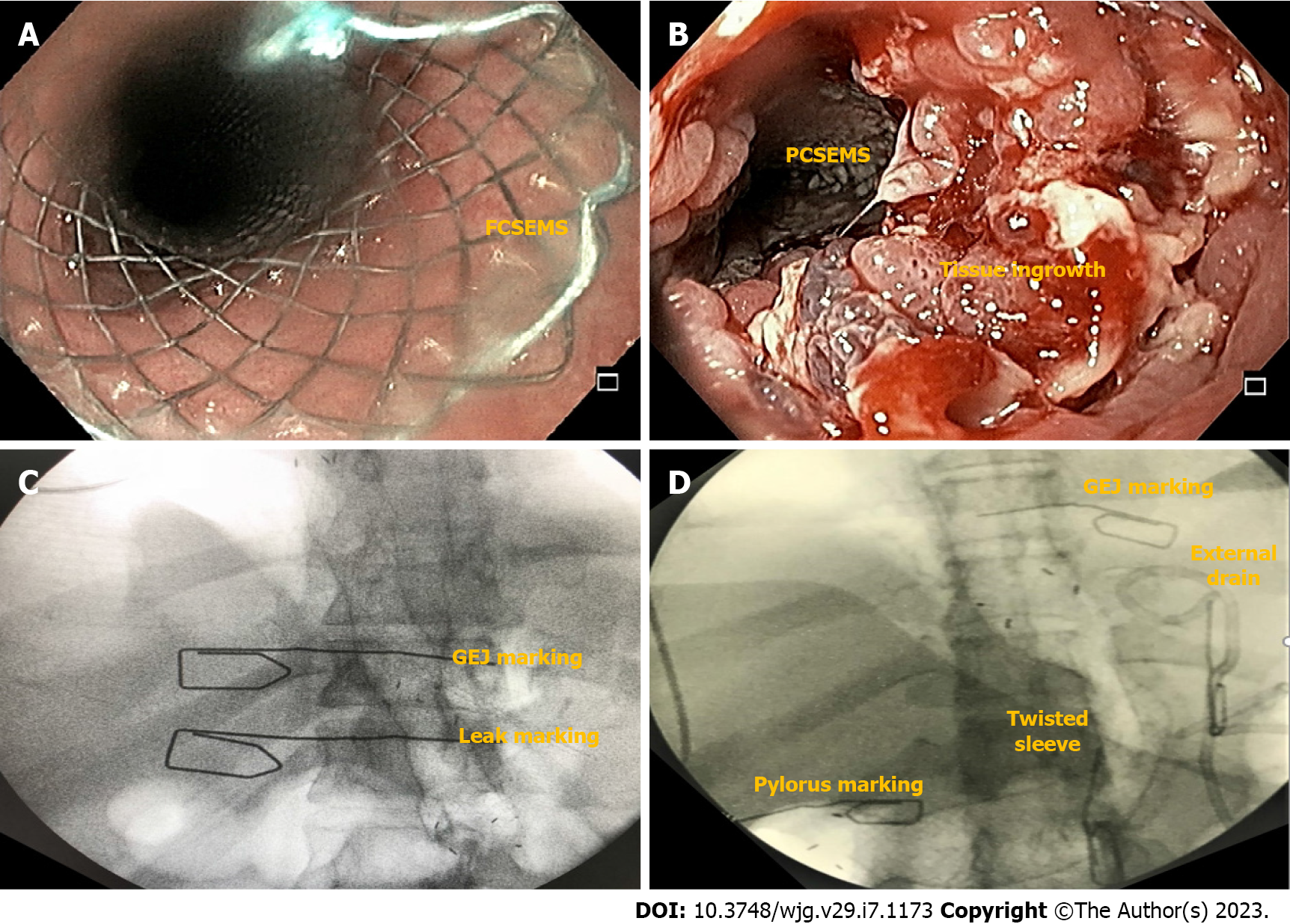
Figure 7 Stents for the treatment of post bariatric and metabolic surgery leaks.
A: Esophageal fully covered self-expandable metal stent; B: Tissue in-growth in an esophageal partially covered self-expandable metal stent; C: Esophageal stent for the treatment of an acute leak after Roux-en-Y gastric bypass; D: Customized bariatric stent for the management of an acute leak in the angle of His after laparoscopic sleeve gastrectomy showing a stenosis at the level of the incisura angularis. FCSEMS: Fully covered self-expandable metal stents; GEJ: Gastroesophageal junction; PCSEMS: Partially covered self-expandable metal stents.
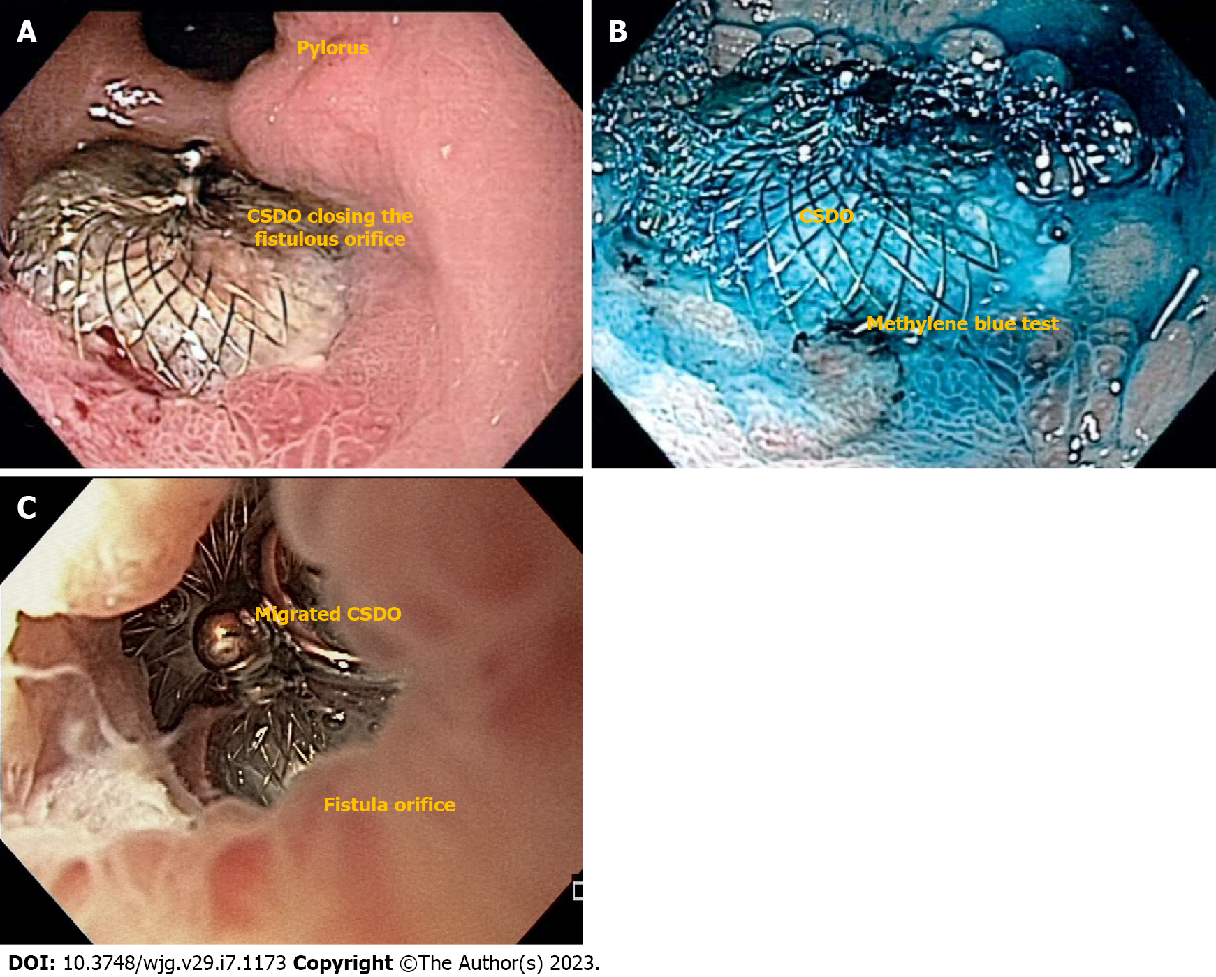
Figure 8 Cardiac septal defect occluder for the treatment of chronic fistulas after bariatric and metabolic surgery.
A: Endoscopic imaging of a successful closure of a gastrocutaneous fistula after cardiac septal defect occluder (CSDO) placement in a distal staple line leak after laparoscopic sleeve gastrectomy; B: Methylene blue test showing no leakage to the skin; C: CSDO migration after placement in an early leak after Roux-en-Y gastric bypass surgery. CSDO: Cardiac septal defect occluder.
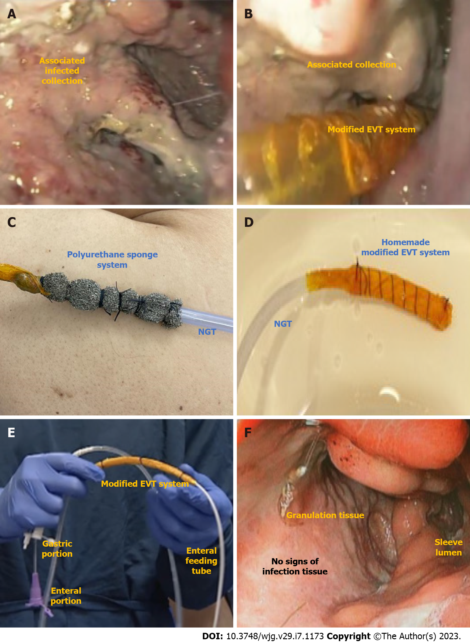
Figure 9 Endoscopic vacuum therapy for the treatment of a post bariatric and metabolic surgery leak.
A: Leak associated collection after laparoscopic sleeve gastrectomy; B: Intracavitary modified endoscopic vacuum therapy (EVT) placement; C: Traditional (polyurethane) sponge system; D: Homemade modified EVT; E: Triple lumen tube used for intraluminal EVT allowing for drainage and nutrition with one tube through the nares; F: Complete tissue healing showing granulation tissue and no signs of infection after EVT treatment. EVT: Endoscopic vacuum therapy; NGT: Nasogastric tube.
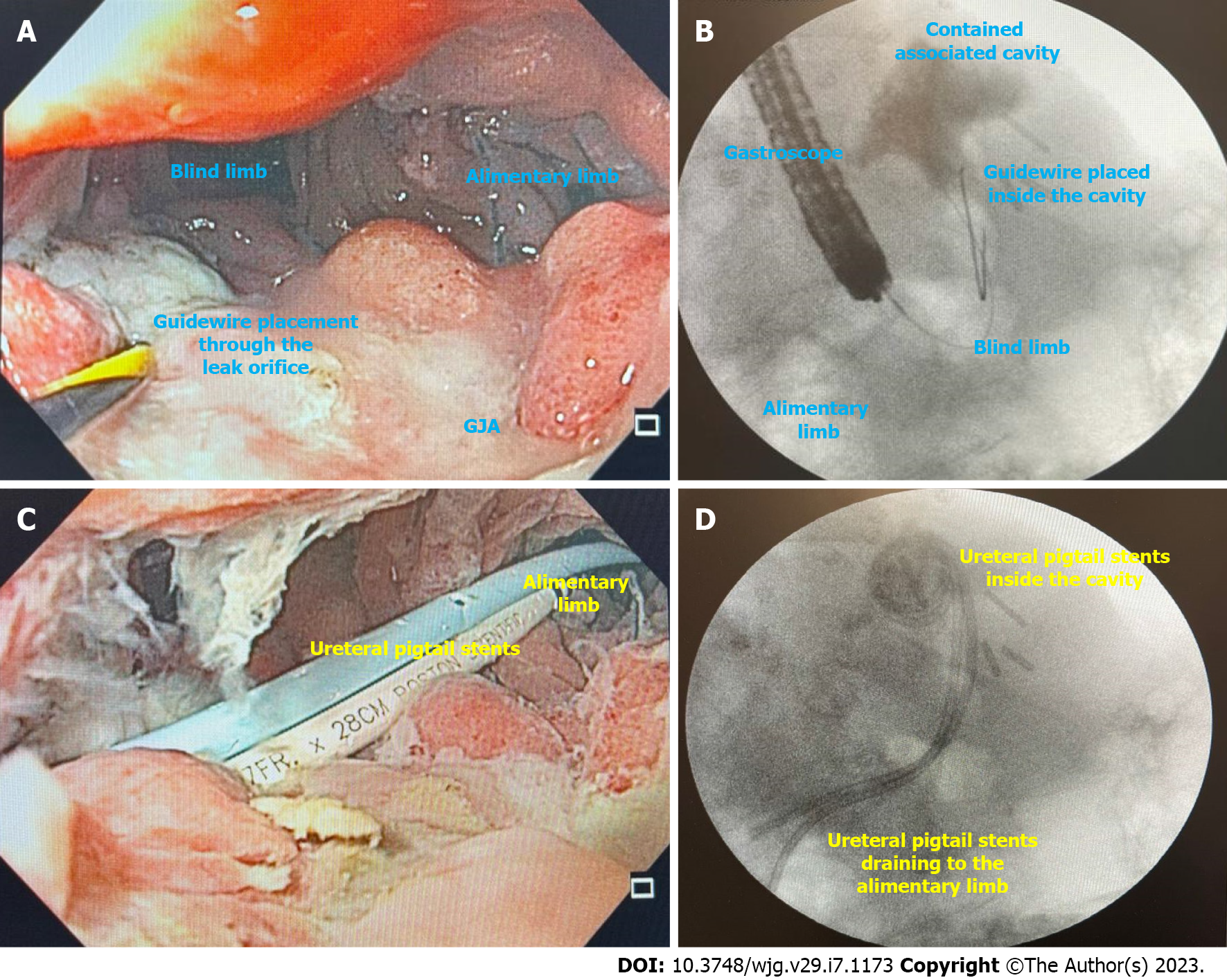
Figure 10 Endoscopic internal drainage with double pigtail stents.
A: Leak orifice at the gastrojejunal anastomosis suture line; after Roux-en-Y gastric bypass surgery; B: Fluoroscopy showing a contained associated collection and guidewire placement; C: Endoscopic internal drainage with ureteral pigtail stents; D: Fluoroscopy showing adequate placement and drainage of the leak associated collection. GJA: Gastrojejunal anastomosis.
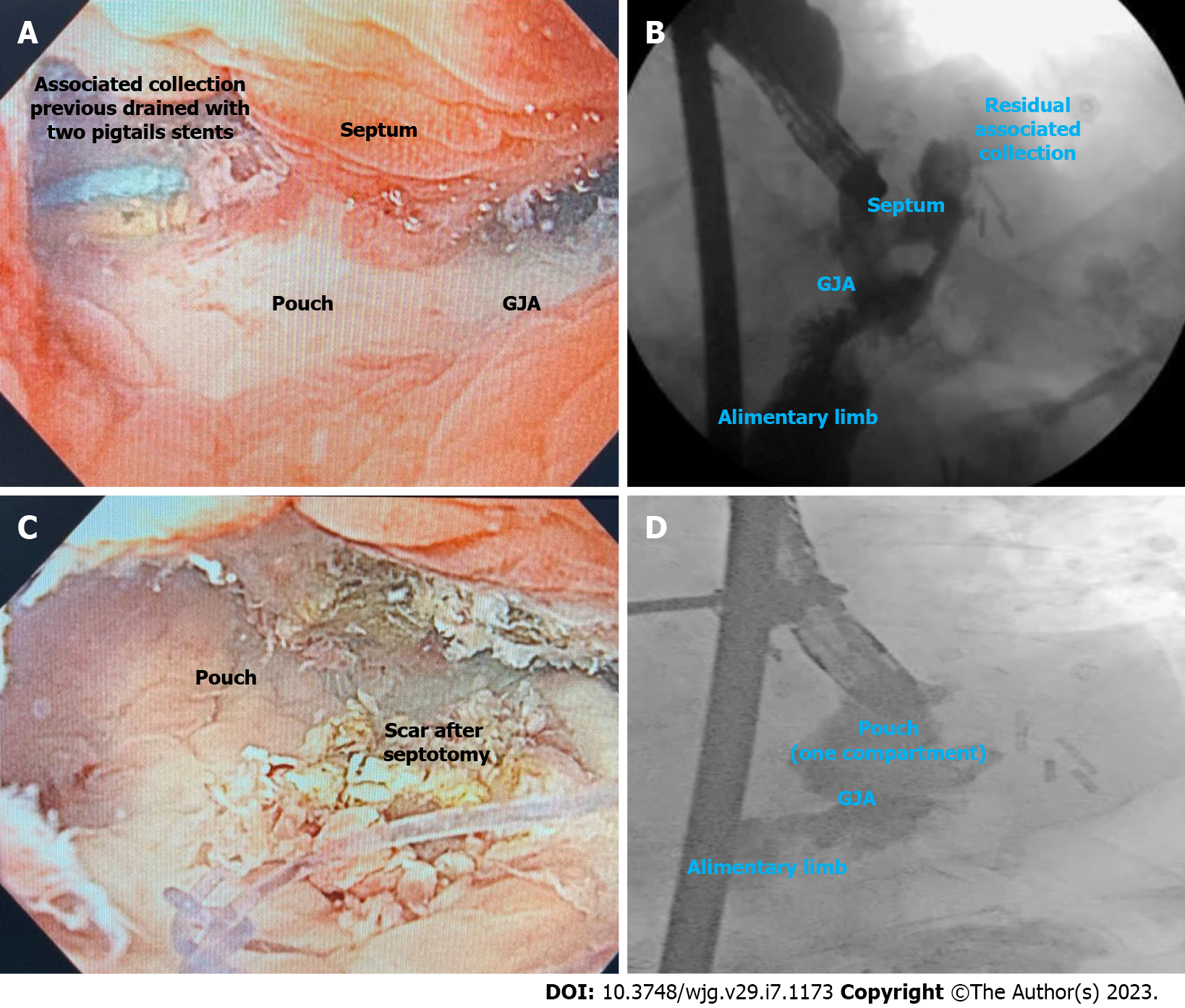
Figure 11 Septotomy.
A: Septum between the associated collection and the leak orifice at the gastrojejunal anastomosis staple line after Roux-en-y gastric bypass surgery; B: Fluoroscopy showing a septum between the leak associated collection and the gastric pouch; C: Endoscopic image after septotomy; D: Fluoroscopy showing a gastric pouch without associated collections as the septotomy transformed the pouch and the associated collection in one compartment. GJA: Gastrojejunal anastomosis.
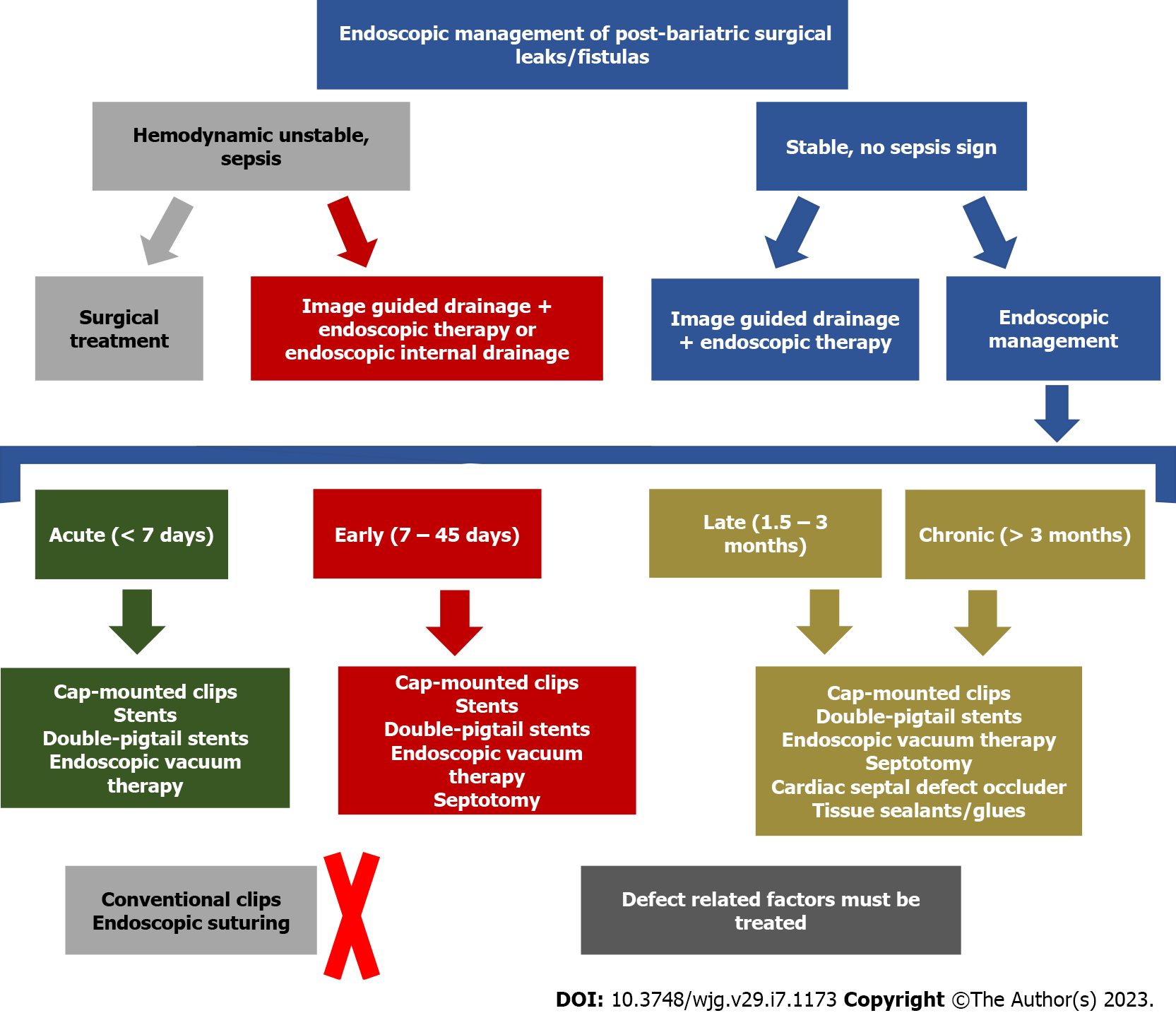
Figure 12 Suggested algorithm for the management of post bariatric surgical leaks and fistulas.
- Citation: de Oliveira VL, Bestetti AM, Trasolini RP, de Moura EGH, de Moura DTH. Choosing the best endoscopic approach for post-bariatric surgical leaks and fistulas: Basic principles and recommendations. World J Gastroenterol 2023; 29(7): 1173-1193
- URL: https://www.wjgnet.com/1007-9327/full/v29/i7/1173.htm
- DOI: https://dx.doi.org/10.3748/wjg.v29.i7.1173


