Copyright
©The Author(s) 2023.
World J Gastroenterol. Feb 7, 2023; 29(5): 834-850
Published online Feb 7, 2023. doi: 10.3748/wjg.v29.i5.834
Published online Feb 7, 2023. doi: 10.3748/wjg.v29.i5.834
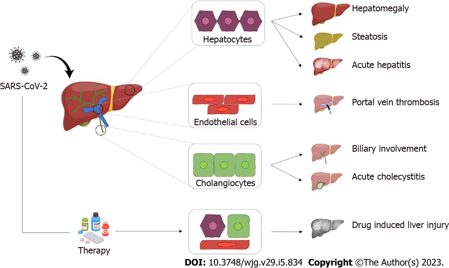
Figure 1 Graphical summary of the most common hepatic pathological findings in coronavirus disease 2019.
SARS-CoV-2: Severe acute respiratory syndrome coronavirus 2.
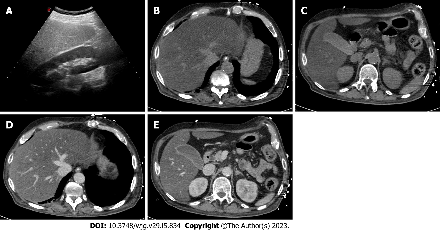
Figure 2 A 45-year-old woman with coronavirus disease 2019 infection underwent abdominal ultrasonography due to elevated liver enzymes.
A: As shown on ultrasonography, the liver is hyperechoic in comparison to the renal parenchyma, suggesting marked steatosis; B-E: The patient underwent contrast-enhanced computed tomography; B and C: On unenhanced images, the liver is diffusely hypoattenuating while its main vessels (i.e. portal vein and hepatic veins) appear brighter than the parenchyma due to a marked hepatic fatty infiltration; D and E: On post-contrast images, the liver is homogeneous without any focal lesion; C and E: The gallbladder is filled by sludge, with normal wall thickness. Finally, the combination of clinical and radiological findings allowed the final diagnosis of steatohepatitis.
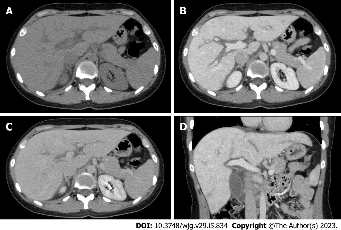
Figure 3 A 33-year-old woman with coronavirus disease 2019 infection underwent contrast-enhanced computed tomography due to a suspicion of portal vein thrombosis.
A: On unenhanced phase liver is enlarged, homogeneous and without any focal lesion; B: On the portal venous phase the liver enhancement is slight inhomogeneous associated with peri-portal edema, a typical finding of acute hepatitis; C and D: These aspects can be clearly observed also on the coronal reconstruction acquired on the portal venous phase (D) and the delayed phase (C).
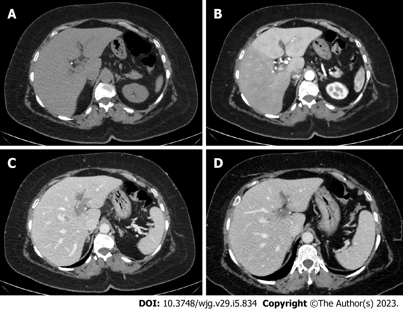
Figure 4 A 40-year-old men with coronavirus disease 2019 infection and marked respiratory symptoms, underwent contrast-enhanced computed tomography due to elevated liver and biliary enzymes.
A and B: On unenhanced phase (A) the liver is within normal limits. After the injection of contrast agent, on arterial phase (B) liver parenchyma shows inhomogeneous enhancement, with hypervascularization of the left liver lobe, as in case of transient hepatic attenuation differences; C: On the portal venous phase, the arterial hypervascularization fades to homogeneous enhancement and diffuse thrombosis of the left branch of the portal vein is demonstrated; D: After 6 mo the patient underwent contrast-enhanced computed tomography that demonstrated persistent portal vein thrombosis without venous collaterals.
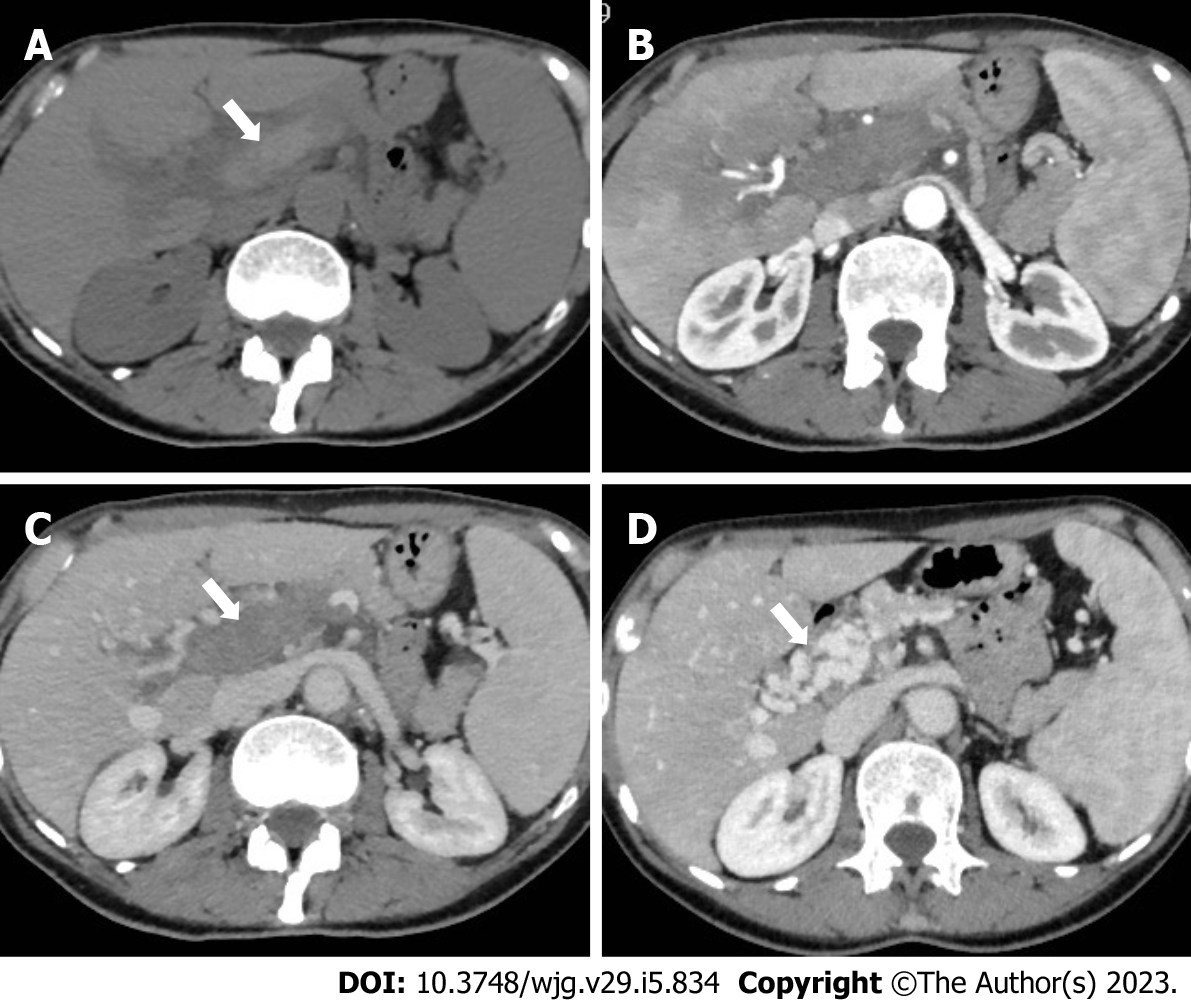
Figure 5 A 62-year-old woman with acute portal vein thrombosis after coronavirus disease 2019 infection.
A-C: Computed tomography (CT) images show acute thrombosis with hyperintense thrombus on unenhanced phase (A, arrow), heterogeneous enhancement of the liver parenchyma on hepatic arterial phase (B), and complete portal vein thrombosis on portal venous phase (C, arrow); D: Contrast-enhanced CT at 6-mo follow-up demonstrates chronic findings of portal cavernoma with multiple collateral vessels at the hepatic hilum (arrow).
Figure 6 A 66-year-old men with coronavirus disease 2019 infection, associated with abdominal pain in the upper quadrants.
A-C: Contrast-enhanced computed tomography showed diffuse thickening of the gallbladder walls (A), with homogeneous contrast enhancement (B), better appreciable on the sagittal reconstruction (C). The gallbladder lumen is filled with biliary sludge (A) without calcified stones. The final diagnosis was acute acalculous cholecystitis.
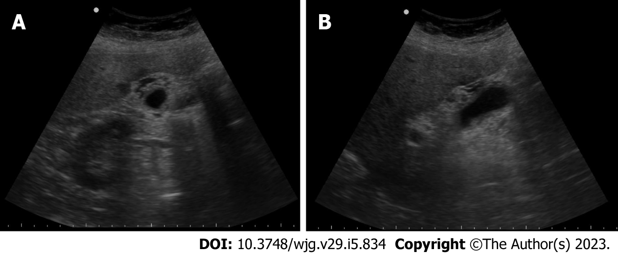
Figure 7 A 53-year-old woman with coronavirus disease 2019 infection and markedly elevated biliary enzymes and C reactive protein levels.
A and B: Ultrasonography demonstrates diffuse thickening of the gallbladder wall, associated with multiple small anechoic components into the walls, as in case of intramural abscesses. No stone is appreciated. The final diagnosis was acute acalculous cholecystitis.
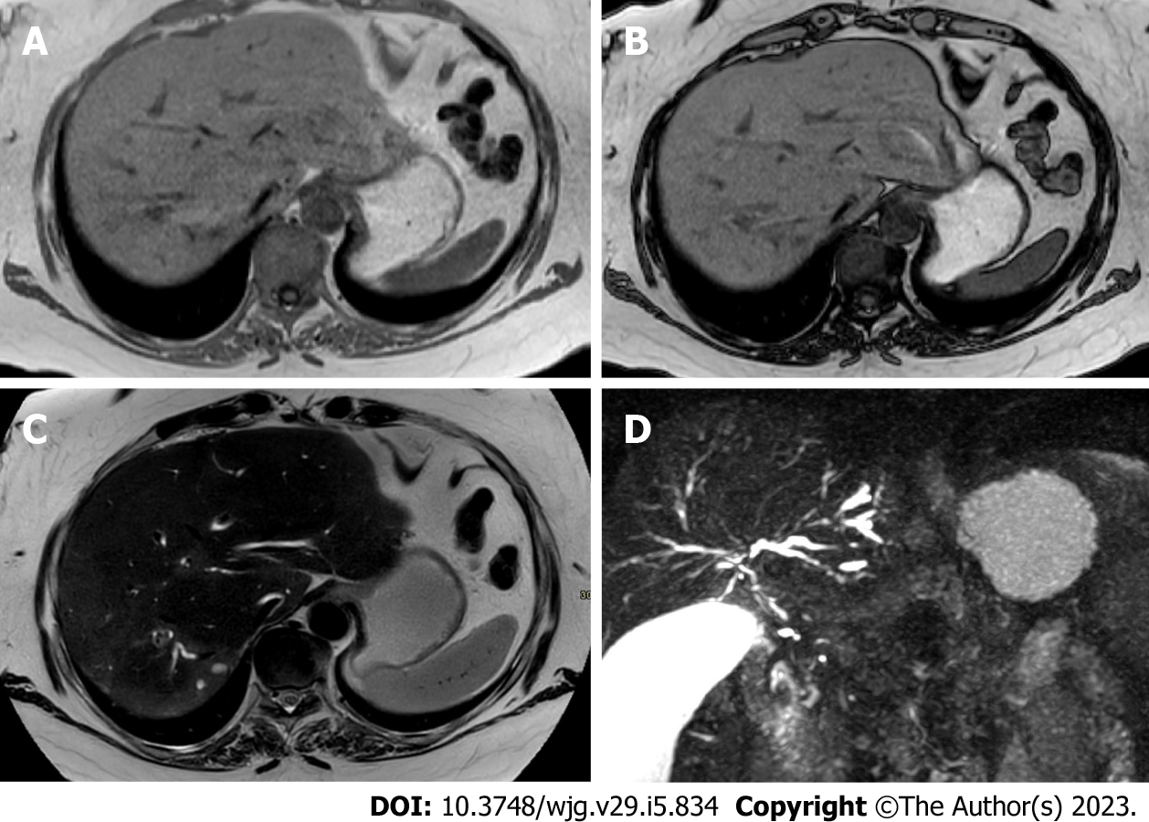
Figure 8 A 44-year-old woman with coronavirus disease 2019 infection underwent magnetic resonance cholangiopancreatography due to elevated biliary enzymes with negative findings on ultrasonography and contrast-enhanced computed tomography.
A and B: In-phase (A) and out-of-phase (B) imaging demonstrates hepatic homogeneous parenchyma without areas of fatty infiltration; C: On the T2-weighted image liver inflammation, especially in the right lobe, is characterized by areas of slight hyperenhancement, located nearby the biliary ducts; D: Finally, magnetic resonance cholangiopancreatography allows the evaluation of the biliary tree, showing multiple and diffuse focal biliary stenosis, in particular in the right lobe, with upstream dilation of small biliary ducts. This radiological aspect is typical of sclerosing cholangitis and the final diagnosis was a biliary involvement due to severe acute respiratory syndrome coronavirus 2.
- Citation: Ippolito D, Maino C, Vernuccio F, Cannella R, Inchingolo R, Dezio M, Faletti R, Bonaffini PA, Gatti M, Sironi S. Liver involvement in patients with COVID-19 infection: A comprehensive overview of diagnostic imaging features. World J Gastroenterol 2023; 29(5): 834-850
- URL: https://www.wjgnet.com/1007-9327/full/v29/i5/834.htm
- DOI: https://dx.doi.org/10.3748/wjg.v29.i5.834


