Copyright
©The Author(s) 2023.
World J Gastroenterol. Dec 28, 2023; 29(48): 6198-6207
Published online Dec 28, 2023. doi: 10.3748/wjg.v29.i48.6198
Published online Dec 28, 2023. doi: 10.3748/wjg.v29.i48.6198
Figure 1 Flow chart of study design and data acquisition.
BE: Barrett’s esophagus; GERD: Gastroesophageal reflux disease; CSMUH: Chung Shan Medical University Hospital; CCH: Changhua Christian Hospital.
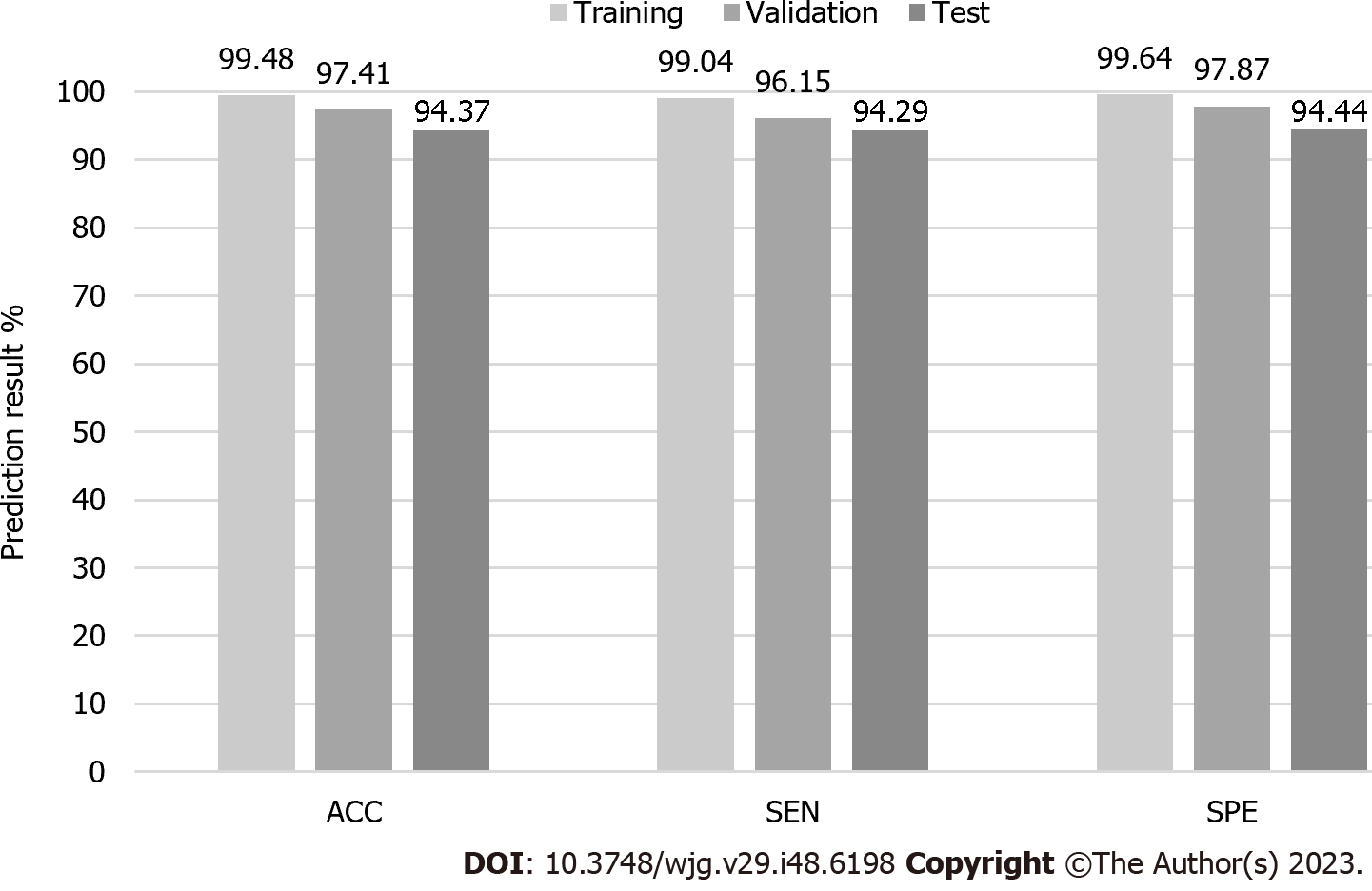
Figure 2 Artificial intelligence image-based Barrett’s esophagus prediction model performance.
ACC: Accuracy; SEN: Sensitivity; SPE: Specificity.
Figure 3 Narrow-band images of the artificial intelligence prediction system.
A: Barrett’s esophagus with successful detection; B: Normal esophagus with successful detection; C: Barrett’s esophagus with false detection; D: Normal esophagus with false detection.
- Citation: Tsai MC, Yen HH, Tsai HY, Huang YK, Luo YS, Kornelius E, Sung WW, Lin CC, Tseng MH, Wang CC. Artificial intelligence system for the detection of Barrett’s esophagus. World J Gastroenterol 2023; 29(48): 6198-6207
- URL: https://www.wjgnet.com/1007-9327/full/v29/i48/6198.htm
- DOI: https://dx.doi.org/10.3748/wjg.v29.i48.6198


