Copyright
©The Author(s) 2023.
World J Gastroenterol. Dec 7, 2023; 29(45): 5988-6016
Published online Dec 7, 2023. doi: 10.3748/wjg.v29.i45.5988
Published online Dec 7, 2023. doi: 10.3748/wjg.v29.i45.5988
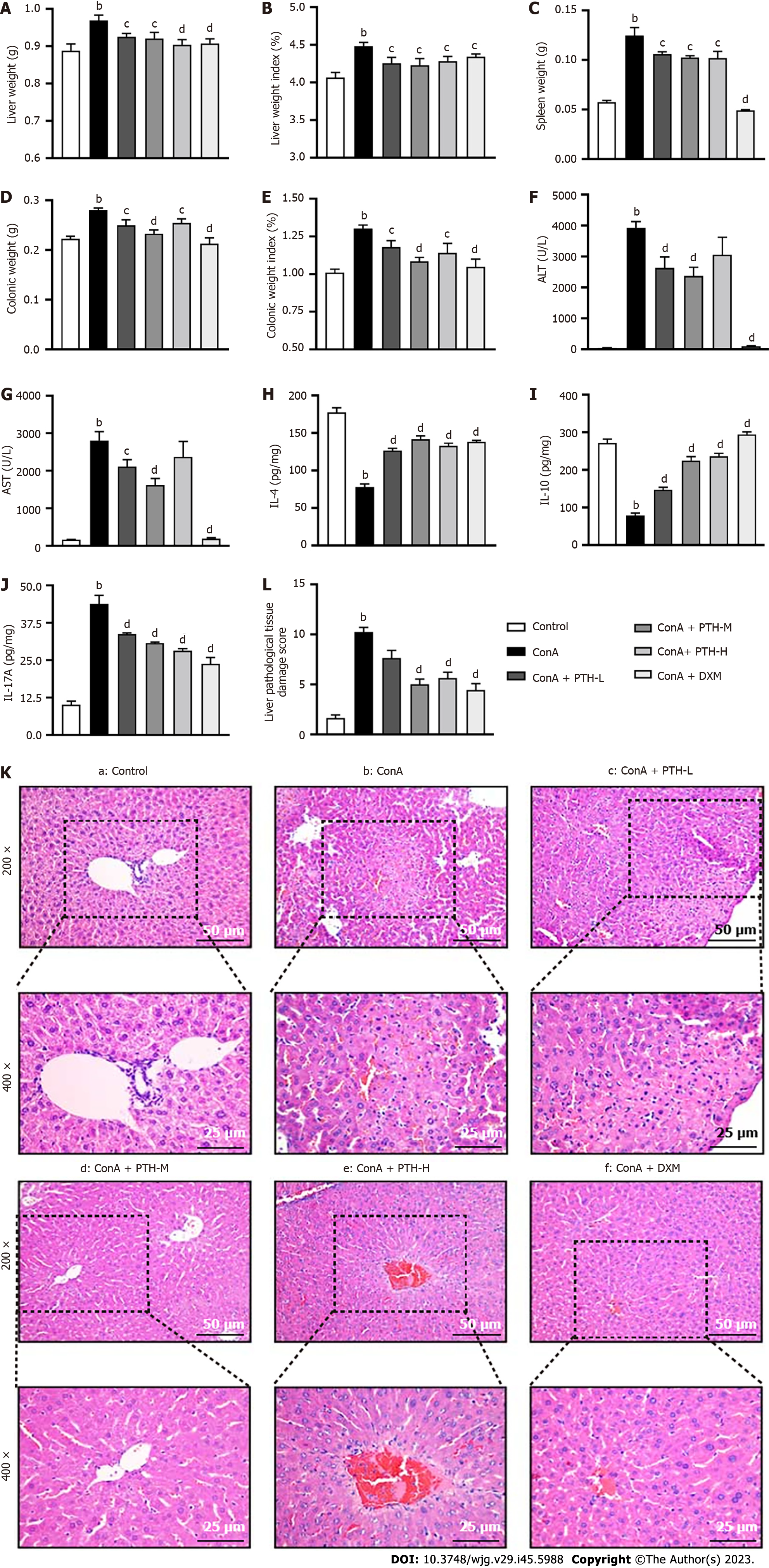
Figure 1 Dose screening of Pien Tze Huang relieved liver injury induced by Concanavalin A in mice.
A: Liver weigh; B: Liver weigh index; C: Spleen weigh; D: Colonic weigh; E: Colonic weigh index; F: Serum alanine aminotransferase; G: Serum aspartate aminotransferase; H-J: The levels of cytokines: Interleukin (IL)-4 (H), IL-10 (I), IL-17A (J) were measured by enzyme-linked immunosorbent assay; K: Representative photographs of hematoxylin and eosin-stained liver sections; L: Scores for liver injury in different groups. According to the Ishak scoring system, liver injury was scored with respect to portal and intralobular inflammation, fragmented necrosis, and bridging necrosis. The extent of the pathology was scored from 0 (no pathology) to 4 (severe pathology). Data are representative images or the mean ± SEM (n = 8). aP < 0.05 and bP < 0.01 vs the control group; cP < 0.05 and dP < 0.01 vs the Concanavalin A group. ALT: Alanine aminotransferase; AST: Aspartate aminotransferase; IL: Interleukin; ConA: Concanavalin A; PTH: Pien Tze Huang; DXM: Dexamethasone.
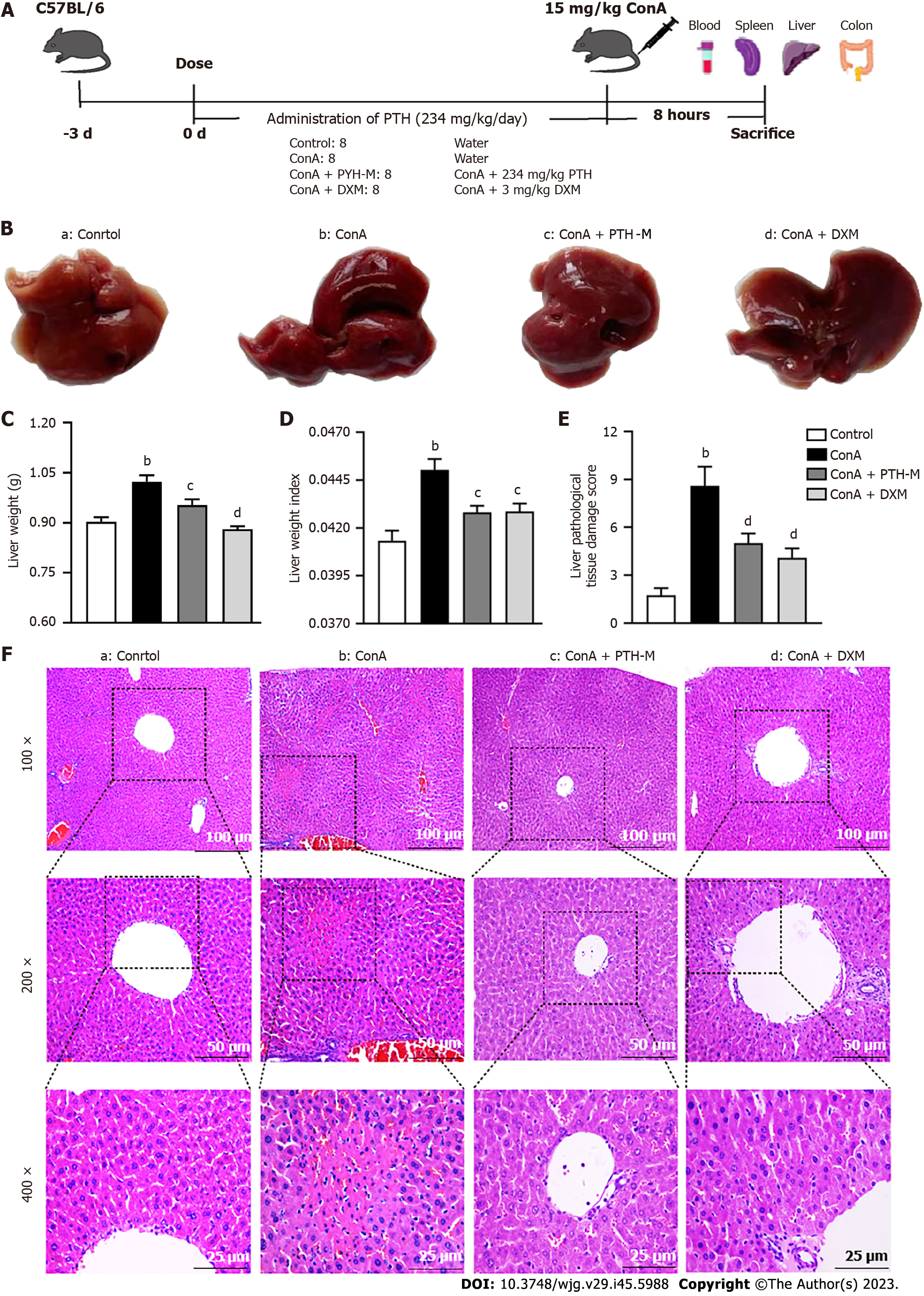
Figure 2 Pien Tze Huang relieves liver injury induced by Concanavalin A in mice.
A: Timeline of the experimental process of treatment and autoimmune hepatitis induction in mice; B-E: Images of mouse livers (B), liver weigh (C), liver weigh index (D), and scores for liver injury (E) in different groups. According to the Ishak scoring system, liver injury was scored with respect to portal and intralobular inflammation, fragmented necrosis, and bridging necrosis. The extent of the pathology was scored from 0 (no pathology) to 4 (severe pathology); F: Representative photographs of hematoxylin and eosin-stained liver sections. Data are representative images or the mean ± SEM (n = 8). aP < 0.05 and bP < 0.01 vs the control group; cP < 0.05 and dP < 0.01 vs the Concanavalin A group. ConA: Concanavalin A; PTH: Pien Tze Huang; DXM: Dexamethasone.
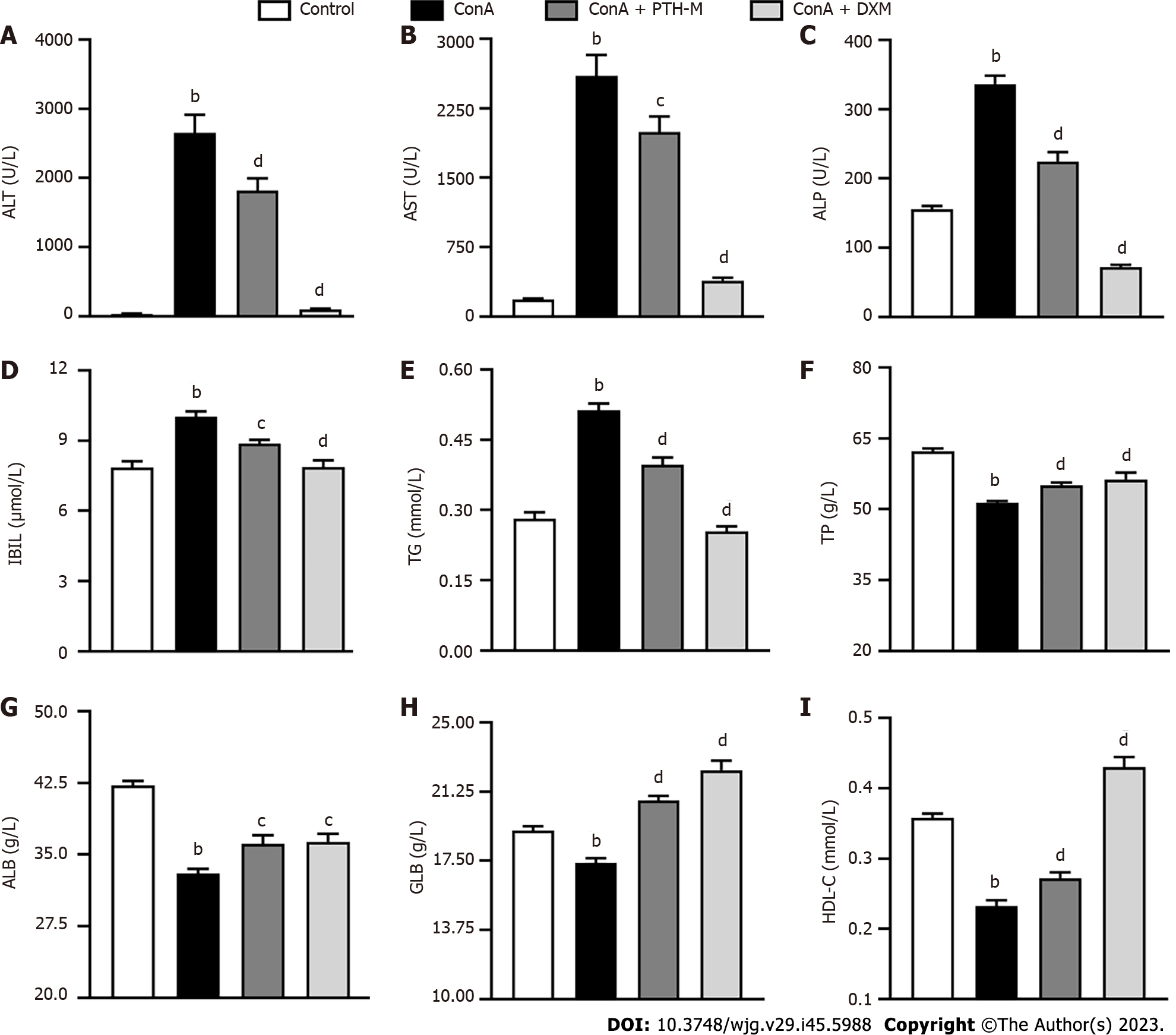
Figure 3 Pien Tze Huang improves liver function in mice with autoimmune hepatitis.
A: Serum alanine aminotransferase; B: Serum aspartate aminotransferase; C: Serum alkaline phosphatase; D: Serum indirect bilirubin; E: Serum triglyceride; F: Serum total protein; G: Serum albumin; H: Serum globulin; I: Serum high-density lipoprotein. Data are representative images or the mean ± SEM (n = 8). aP < 0.05 and bP < 0.01 vs the control group; cP < 0.05 and dP < 0.01 vs the Concanavalin A group. ALT: Alanine aminotransferase; AST: Aspartate aminotransferase; ALP: Alkaline phosphatase; IBIL: Indirect bilirubin; TG: Triglyceride; TP: Total protein; ALB: Albumin; GLB: Globulin; HDL: High-density lipoprotein; ConA: Concanavalin A; PTH: Pien Tze Huang; DXM: Dexamethasone.
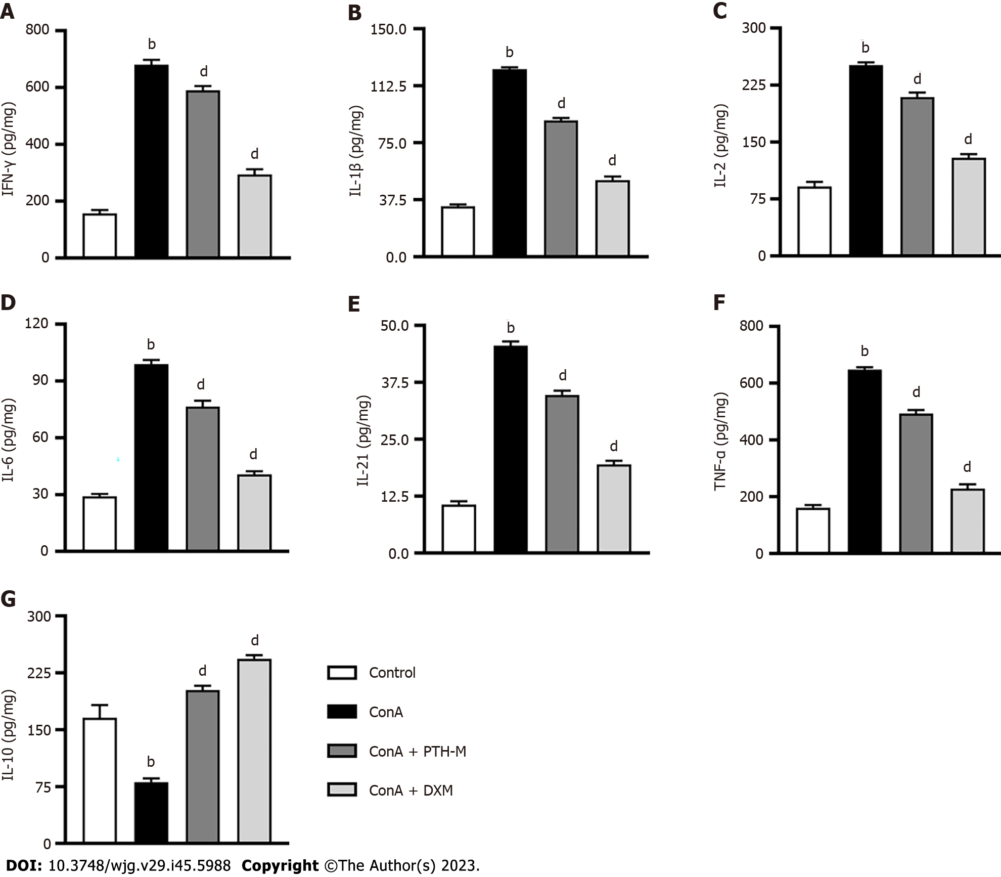
Figure 4 Pien Tze Huang regulates the expression of inflammatory cytokines in mice with autoimmune hepatitis.
A-G: The levels of cytokines: Interferon-γ (A), interleukin (IL)-1β (B), IL-2 (C), IL-6 (D), IL-21 (E), tumor necrosis factor-alpha (F), and IL-10 (G), were measured by enzyme-linked immunosorbent assay. Data are representative images or the mean ± SEM (n = 8). aP < 0.05 and bP < 0.01 vs the control group; cP < 0.05 and dP < 0.01 vs the Concanavalin A group. IFN: Interferon; IL: Interleukin; TNF: Tumor necrosis factor; ConA: Concanavalin A; PTH: Pien Tze Huang; DXM: Dexamethasone.
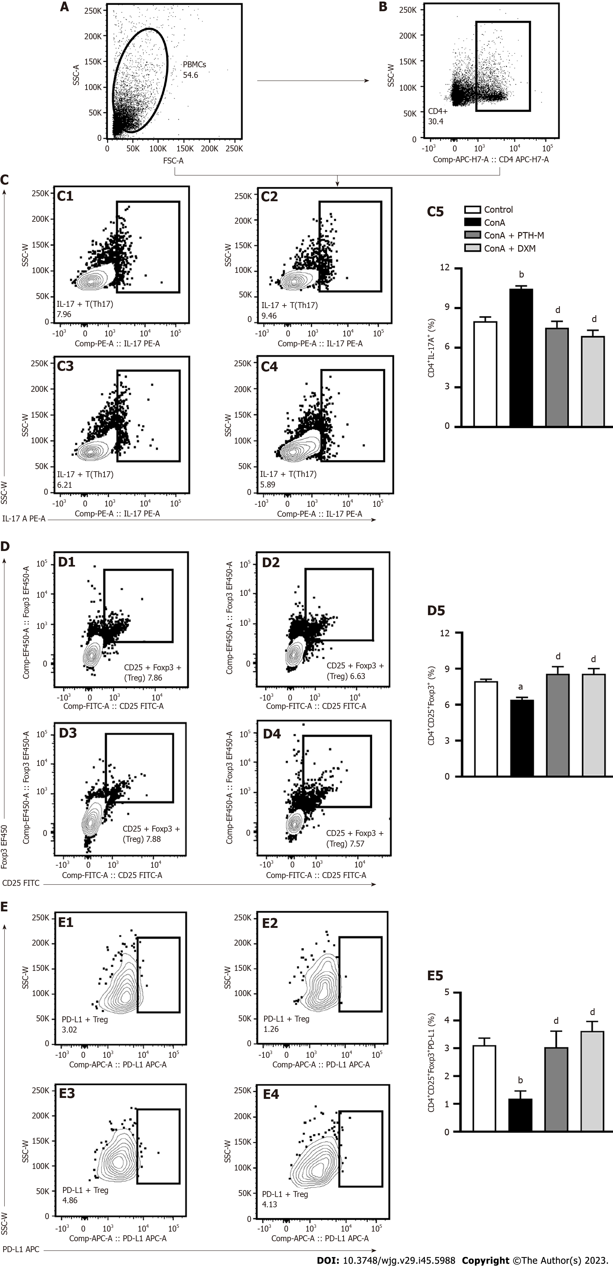
Figure 5 Pien Tze Huang regulates the number of T helper type 17 and regulatory T cells in mice with autoimmune hepatitis.
A: Mononuclear cells in peripheral blood; B: CD4+ lymphocytes measured by flow cytometry; C: CD4+IL-17A+(T helper type 17) cells. C1-C4: Control, Concanavalin A (Con A), Con A + middle-dose Pien Tze Huang (PTH-M), and Con A + dexamethasone (DXM) group, respectively; C5: Statistical analysis of CD4+IL17A+ cell frequencies in these four groups; D: CD4+CD25+Foxp3+ cells. D1–D4: Control, Con A, Con A + PTH-M, and Con A + DXM group, respectively; D5: Statistical analysis of CD4+CD25+Foxp3+ cell frequencies in these four groups; E: CD4+CD25+Foxp3+PD-L1+ cells. E1–E4: Control, Con A, Con A + PTH-M, and Con A + DXM group, respectively; E5: Statistical analysis of CD4+CD25+Foxp3+PD-L1+ cell frequencies in these four groups. Data are representative images or the mean ± SEM (n = 8). aP < 0.05 and bP < 0.01 vs the control group; cP < 0.05 and dP < 0.01 vs the Concanavalin A group. IL: Interleukin; ConA: Concanavalin A; PTH: Pien Tze Huang; DXM: Dexamethasone.
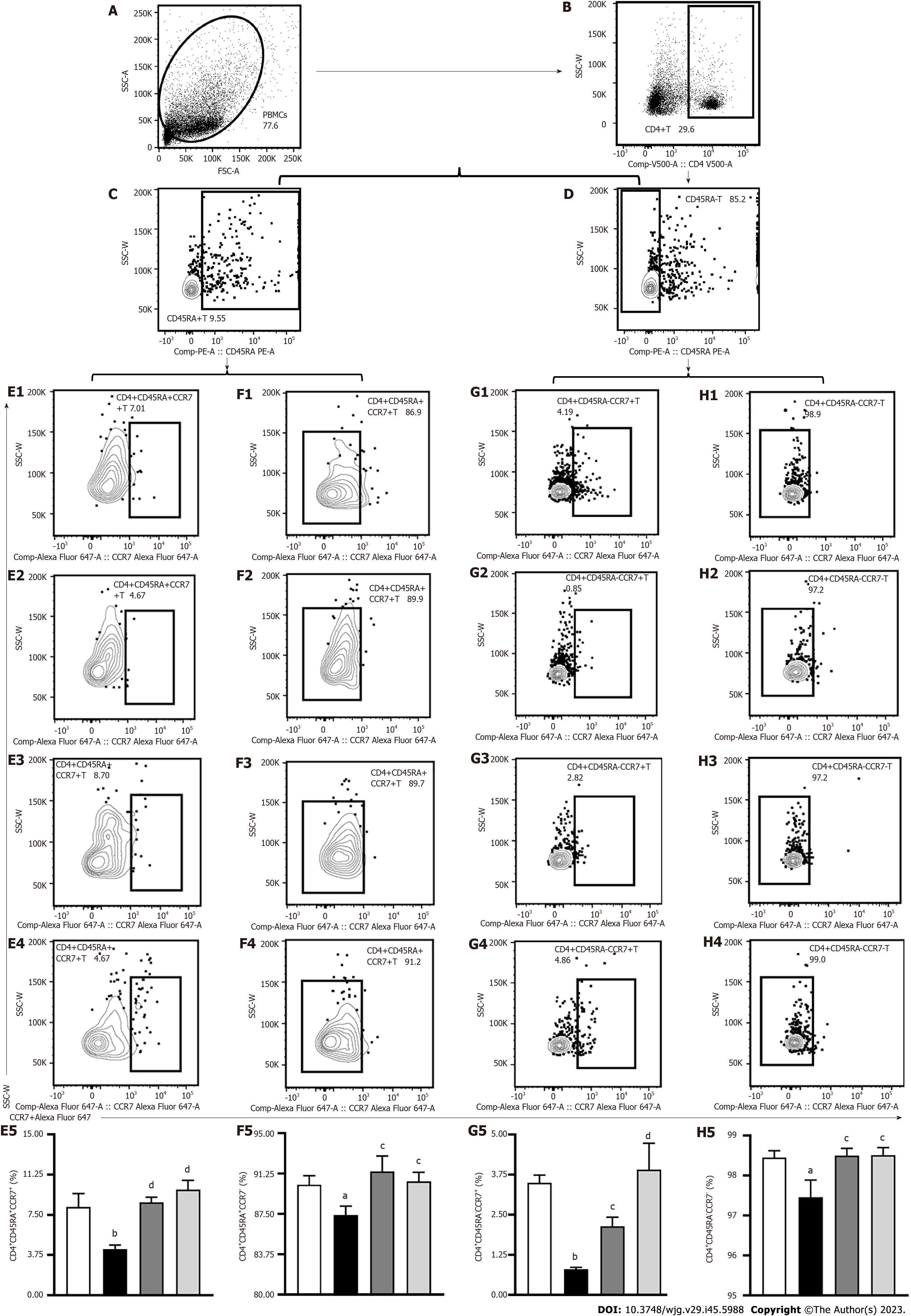
Figure 6 Pien Tze Huang regulates the number of memory T cells in mice with autoimmune hepatitis.
A: Peripheral blood mononuclear cells; B: CD4+ lymphocytes measured by flow cytometry; C: CD45RA+ lymphocytes measured by flow cytometry; D: CD45RA- lymphocytes measured by flow cytometry; E: CD4+CD45RA+CCR7+ cells. E1-E4: Control, Concanavalin A (Con A), Con A + middle-dose Pien Tze Huang (PTH-M), and Con A + dexamethasone (DXM) group in order; E5: Statistical analysis of CD4+CD45RA+CCR7+ cell frequencies in these four groups; F: CD4+CD45RA+CCR7- cells. F1-F4: Control, Con A, Con A + PTH-M, and Con A + DXM group, respectively; F5: Statistical analysis of CD4+CD45RA+CCR7- cell frequencies in these four groups; G: CD4+CD45RA-CCR7+ cells. G1-G4: Control, Con A, Con A + PTH-M, and Con A + DXM group, respectively; G5: Statistical analysis of CD4+CD45RA-CCR7+ cell frequencies in these four groups; H: CD4+CD45RA-CCR7-cells. H1-H4: Control, Con A, Con A + PTH-M, and Con A + DXM group, respectively; H5: Statistical analysis of CD4+CD45RA-CCR7- cell frequencies in these four groups. Data are representative images or the mean ± SEM (n = 8). aP < 0.05 and bP < 0.01 vs the control group; cP < 0.05 and dP < 0.01 vs the Concanavalin A group. ConA: Concanavalin A; PTH: Pien Tze Huang; DXM: Dexamethasone.
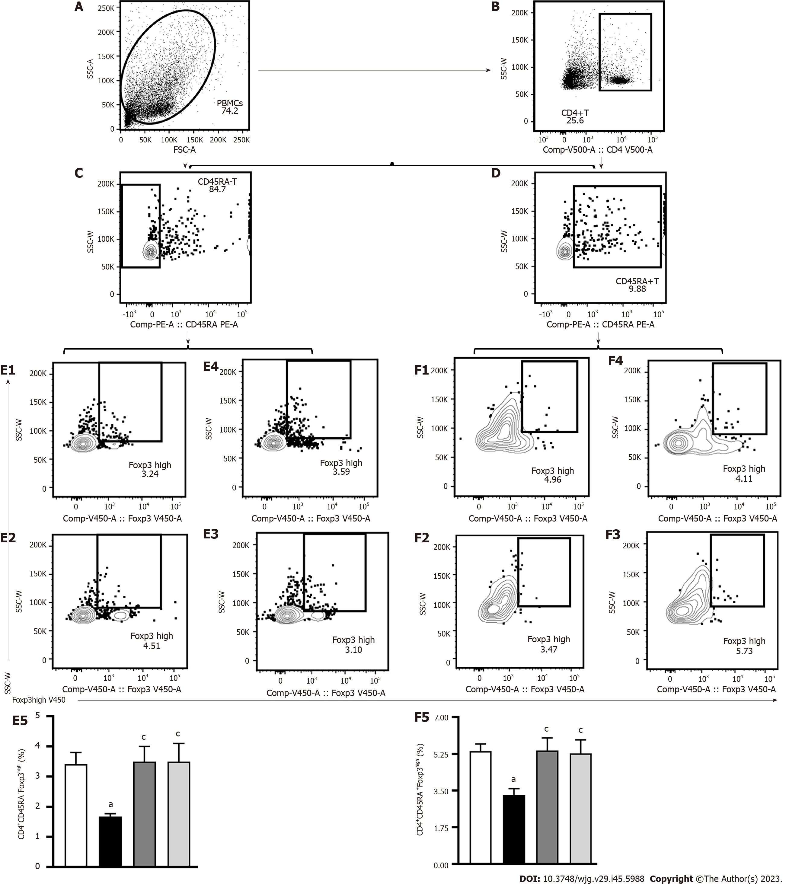
Figure 7 Pien Tze Huang regulates the number of special memory regulatory T cells in mice with autoimmune hepatitis.
A: Peripheral blood mononuclear cells; B: CD4+ lymphocytes measured by flow cytometry; C: CD45RA-lymphocytes measured by flow cytometry; D: CD45RA+ lymphocytes measured by flow cytometry; E: CD4+CD45RA-Foxp3high cells. E1-E4: Control, Concanavalin A (Con A), Con A + middle-dose Pien Tze Huang (PTH-M), and Con A + dexamethasone (DXM) group, respectively; E5: Statistical analysis of CD4+CD45RA-Foxp3high cell frequencies in these four groups; F: CD4+CD45RA+Foxp3high cells. F1-F4: Control, Con A, Con A + PTH-M, and Con A + DXM group, respectively; F5: Statistical analysis of CD4+CD45RA+Foxp3high cell frequencies in these four groups. Data are representative images or the mean ± SEM (n = 8). aP < 0.05 and bP < 0.01 vs the control group; cP < 0.05 and dP < 0.01 vs the Concanavalin A group. ConA: Concanavalin A; PTH: Pien Tze Huang; DXM: Dexamethasone.
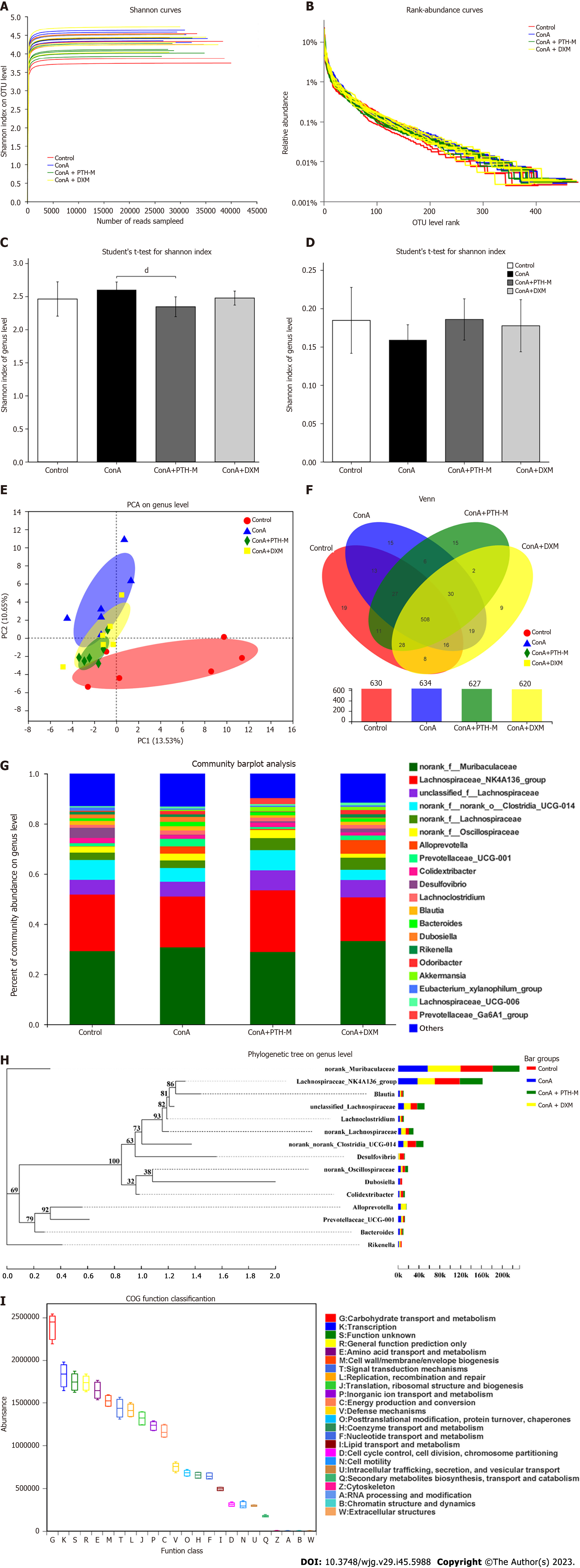
Figure 8 Pien Tze Huang improves the composition of gut microbiota in mice with autoimmune hepatitis.
A: The rarefaction curve of the Shannon index at the operational taxonomic unit (OTU) level; B: Rank-Abundance curves at the OTU level; C: Shannon index at the genus level; D: Simpson index at the genus level; E: Partial least squares discriminant analysis score at the genus level; F: The Venn diagram depicts OTUs that differed in each group; G: Community composition bar chart at the genus level; H: Phylogenetic evolutionary tree at the genus level; I: COG function classification. Data are representative images or the mean ± SEM (n = 6). aP < 0.05 and bP < 0.01 vs the control group; cP < 0.05 and dP < 0.01 vs the Concanavalin A group. ConA: Concanavalin A; PTH: Pien Tze Huang; DXM: Dexamethasone.
Figure 9 Species differences in intestinal microbiota and correlation analysis.
A: Differential analysis among the four groups at the genus level; B: Phylotypes significantly different between the control and Concanavalin A (Con A) groups at the genus level; C: Phylotypes significantly different between Con A and Con A + middle-dose Pien Tze Huang groups at the genus level; D: Redundancy analysis of relationship between toll-like receptor (TLR)4, aspartate aminotransferase (AST), alanine aminotransferase (ALT), HAI, CD4+CD25+Foxp3+, CD4+CD45RA-Foxp3high and intestinal microbiota; E: Distance-based redundancy analysis of relationship between TLR4, AST, ALT, HAI, CD4+CD25+Foxp3+(regulatory T cells) cells, CD4+CD45RA-Foxp3high (memory regulatory T cells) cells, and intestinal microbiota; F: Spearman’s correlation heatmap of TLR4, AST, ALT, HAI, CD4+CD25+Foxp3+, CD4+CD45RA-Foxp3high and intestinal microbiota; G: The quantitative analysis of TLR4 protein expression. Data are representative images or the mean ± SEM (n = 6). aP < 0.05 and bP < 0.01 vs the control group; cP < 0.05 and dP < 0.01 vs the Concanavalin A group. ConA: Concanavalin A; PTH: Pien Tze Huang; DXM: Dexamethasone.
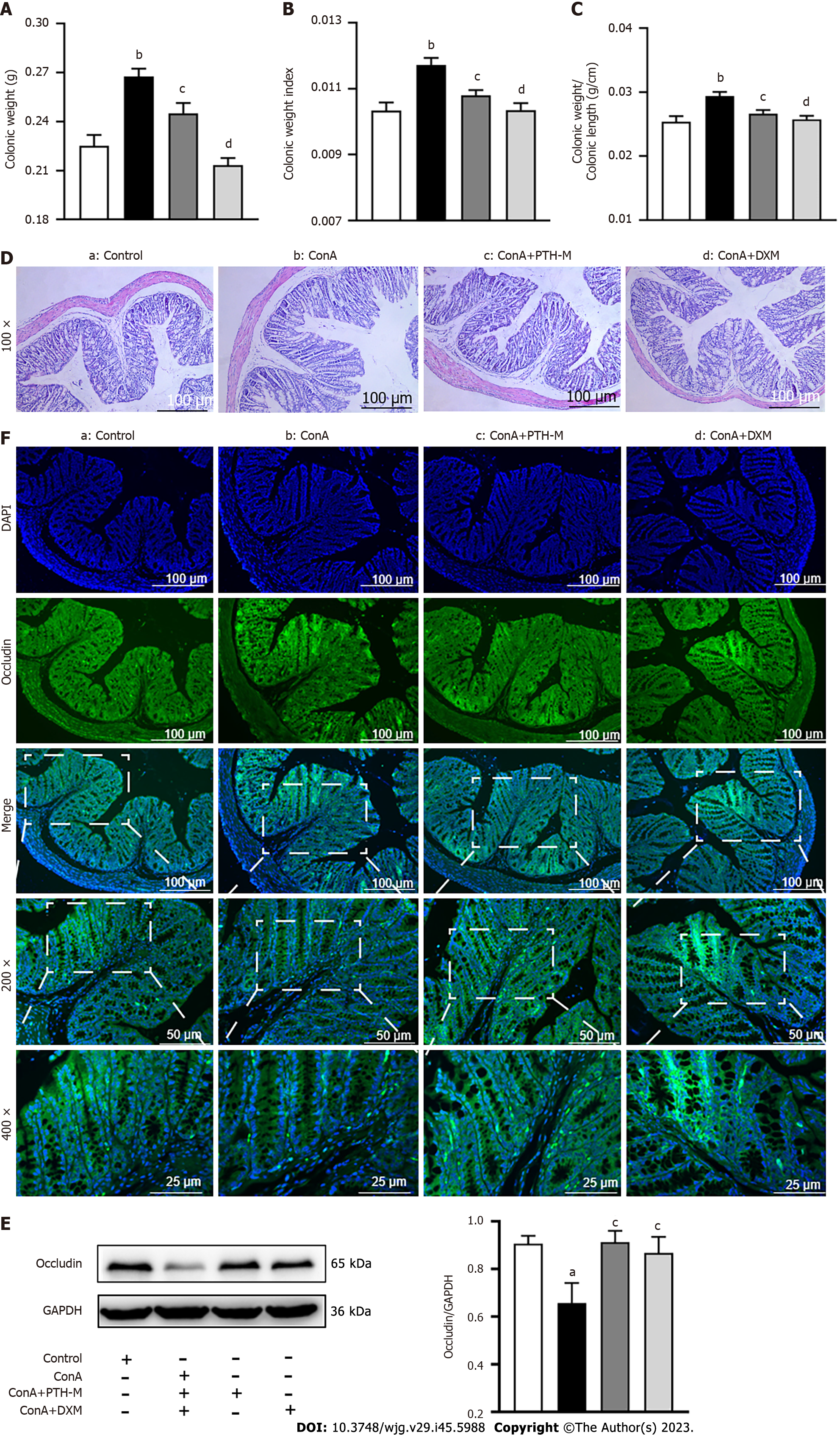
Figure 10 Pien Tze Huang restores the function of the intestinal barrier in mice with autoimmune hepatitis.
A: Colonic weight; B: Colonic weight index; C: Colonic weight/Colonic length; D: Colonic hematoxylin and eosin staining; E: The quantitative analysis of occludin protein expression; F: Representative images of occludin in the colon (IF, 200 ×, 400 ×). Data are representative images or the mean ± SEM (n = 8). aP < 0.05 and bP < 0.01 vs the control group; cP < 0.05 and dP < 0.01 vs the Concanavalin A group. ConA: Concanavalin A; PTH: Pien Tze Huang; DXM: Dexamethasone.
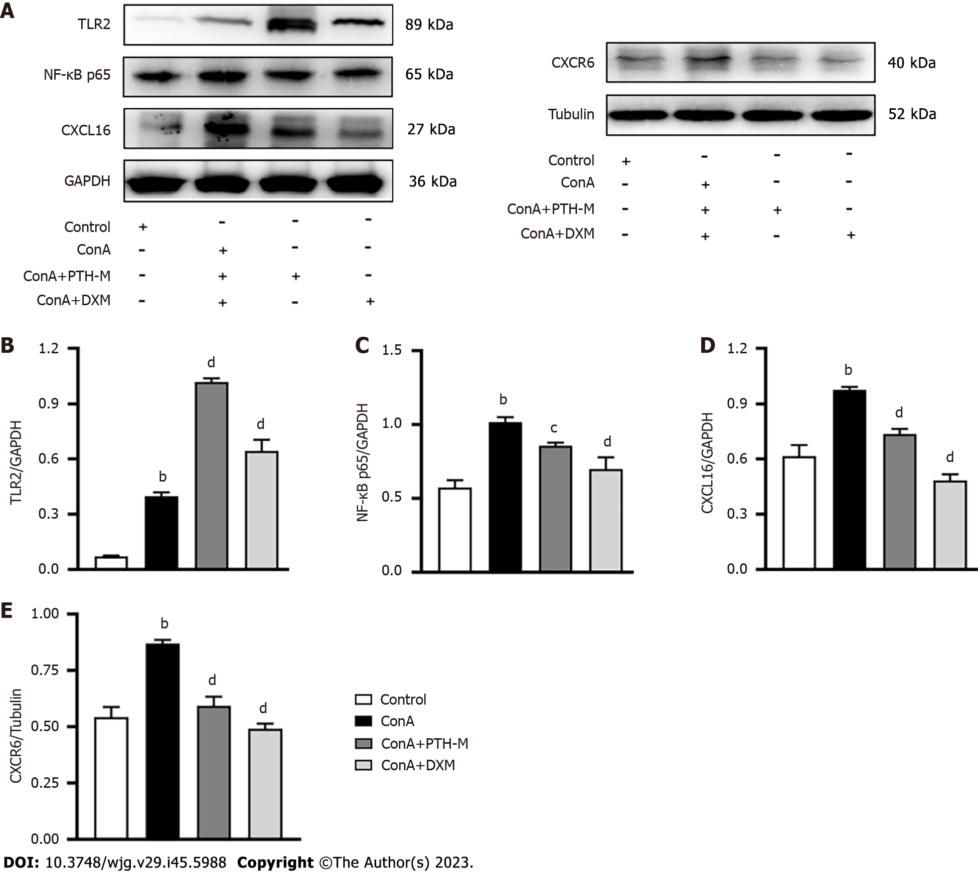
Figure 11 Pien Tze Huang regulates the expression of toll-like receptor 2, nuclear factor-κB p65, CXCR6, and CXCL16 in mice with autoimmune hepatitis.
A: The protein expression of toll-like receptor (TLR)2, nuclear factor-κB (NF-κB) p65, CXCR6, and CXCL16 analyzed by western blot; B: Quantitative analysis of TLR2; C: Quantitative analysis of NF-κB p65; D: Quantitative analysis of CXCL16; E: Quantitative analysis of CXCR6. Data are representative images or the mean ± SEM (n = 8). aP < 0.05 and bP < 0.01 vs the control group; cP < 0.05 and dP < 0.01 vs the Concanavalin A group. TLR: Toll-like receptor; NF-κB: Nuclear factor-κB; ConA: Concanavalin A; PTH: Pien Tze Huang; DXM: Dexamethasone.
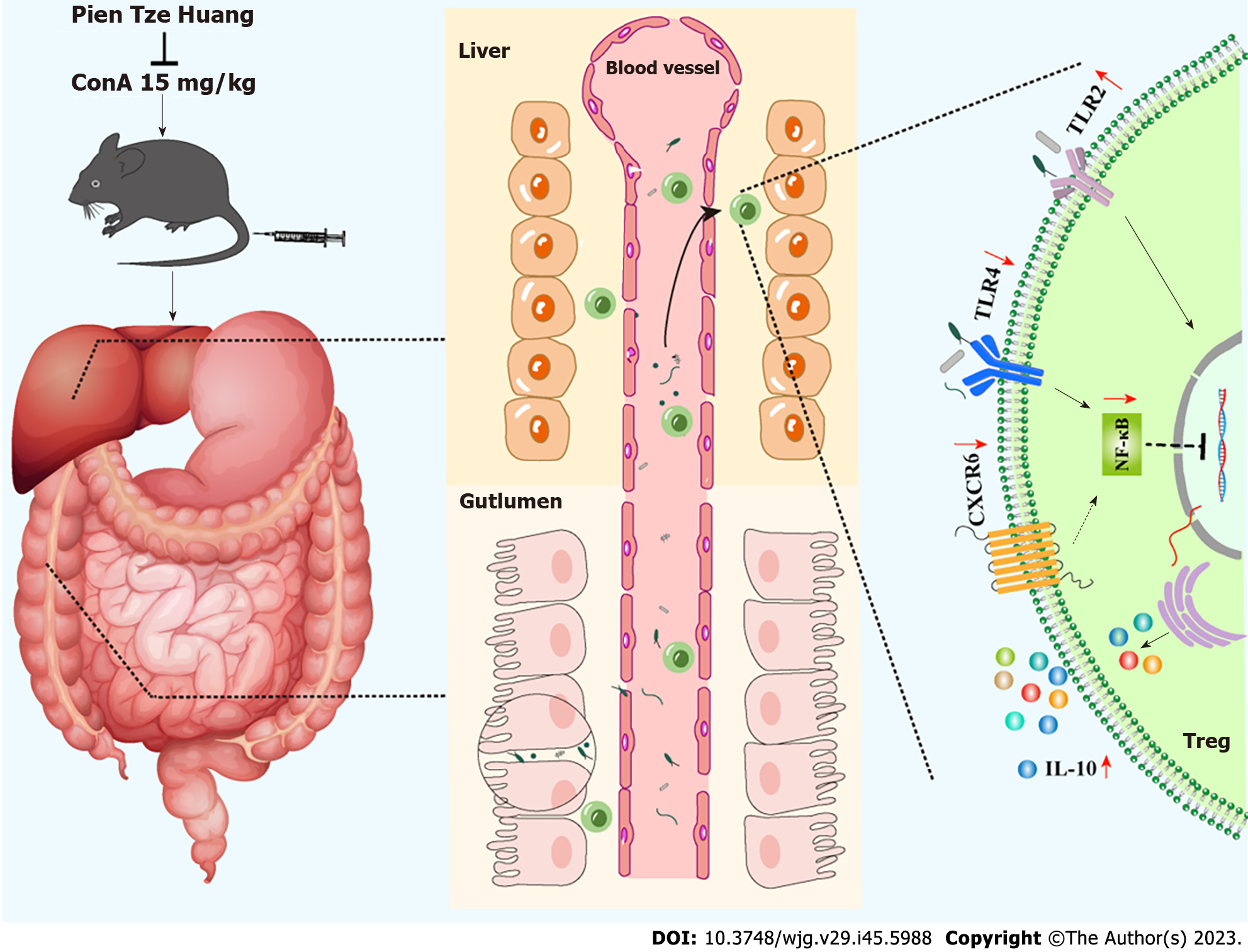
Figure 12 Protective mechanisms of Pien Tze Huang against autoimmune hepatitis.
TLR: Toll-like receptor; NF-κB: Nuclear factor-κB; ConA: Concanavalin A; IL: Interleukin.
- Citation: Zeng X, Liu MH, Xiong Y, Zheng LX, Guo KE, Zhao HM, Yin YT, Liu DY, Zhou BG. Pien Tze Huang alleviates Concanavalin A-induced autoimmune hepatitis by regulating intestinal microbiota and memory regulatory T cells. World J Gastroenterol 2023; 29(45): 5988-6016
- URL: https://www.wjgnet.com/1007-9327/full/v29/i45/5988.htm
- DOI: https://dx.doi.org/10.3748/wjg.v29.i45.5988


