Copyright
©The Author(s) 2023.
World J Gastroenterol. Oct 14, 2023; 29(38): 5361-5373
Published online Oct 14, 2023. doi: 10.3748/wjg.v29.i38.5361
Published online Oct 14, 2023. doi: 10.3748/wjg.v29.i38.5361
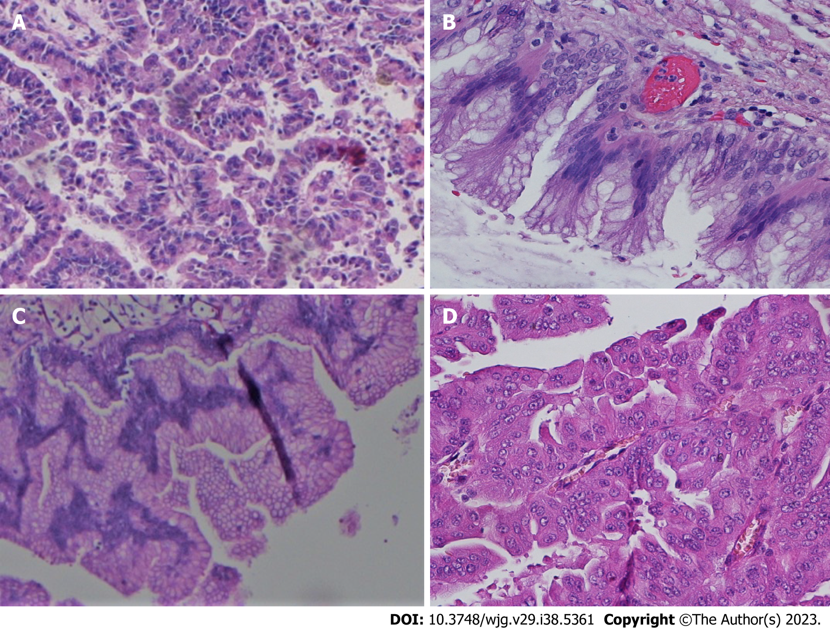
Figure 1 Histological subtypes of intraductal papillary neoplasms of the bile duct.
A: Pancreatobiliary; B: Intestinal; C: Gastric; D: Oncocytic. Hematoxylin and eosin staining.
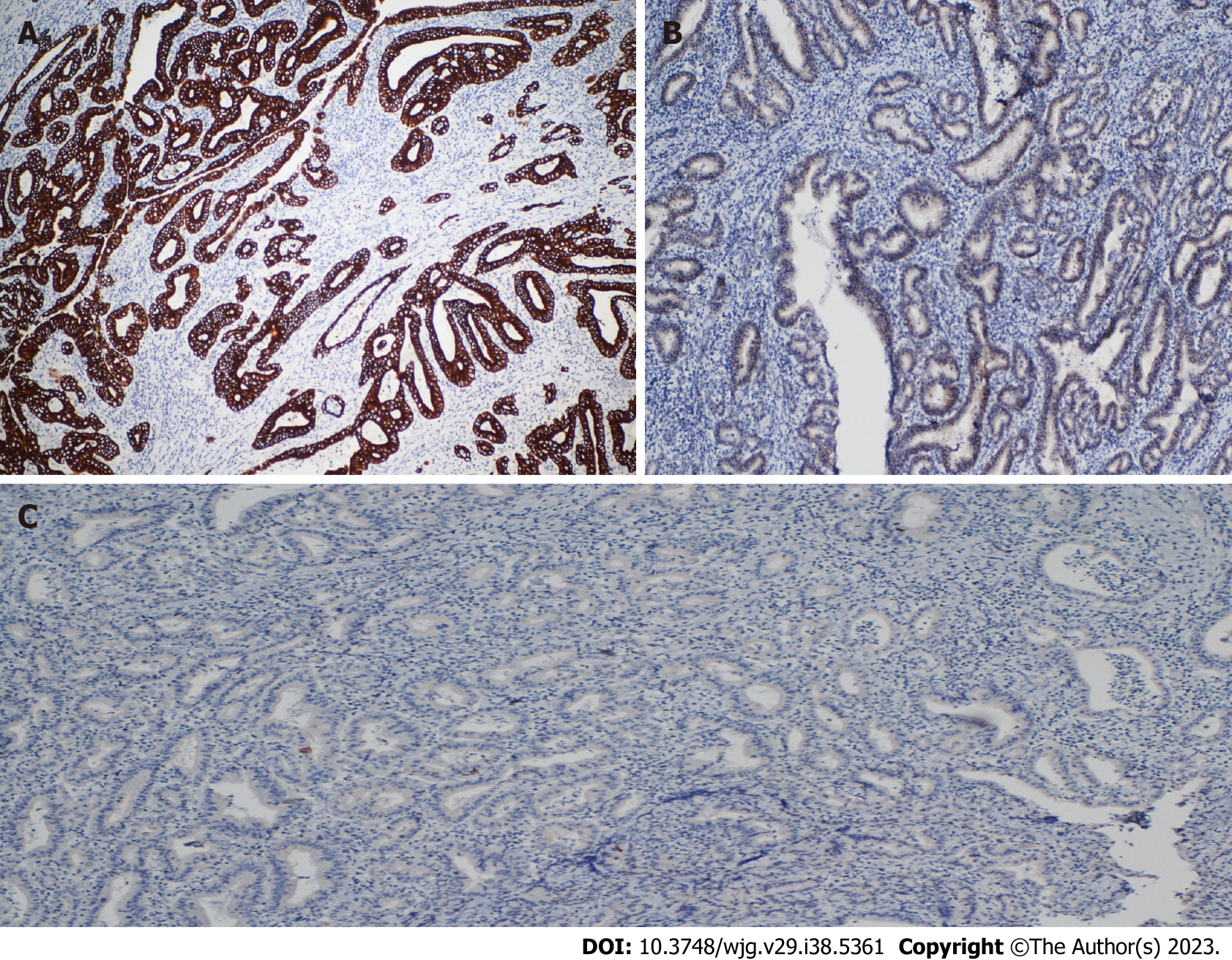
Figure 2 Histological features of intraductal papillary neoplasms of the bile duct with irregular growth pattern and foci of invasive carcinoma (pancreatobiliary type).
A: Immunostaining positive for cytokeratin (CK)19; B: Immunostaining negative for CDX2; C: Immunostaining negative for CK20.
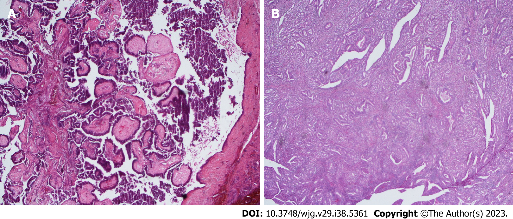
Figure 3 Subclassification of intraductal papillary neoplasms of the bile duct according to the Japan Biliary Association and the Korean Association of Hepato-Biliary-Pancreatic Surgery.
A: Type 1 consists of papillary, villous or tubular homogenous structures with thin papillary fibrovascular stalks; B: Type 2 consists of thick papillary glands with irregular branching, often intermingled with solid irregular components. Hematoxylin and eosin staining.
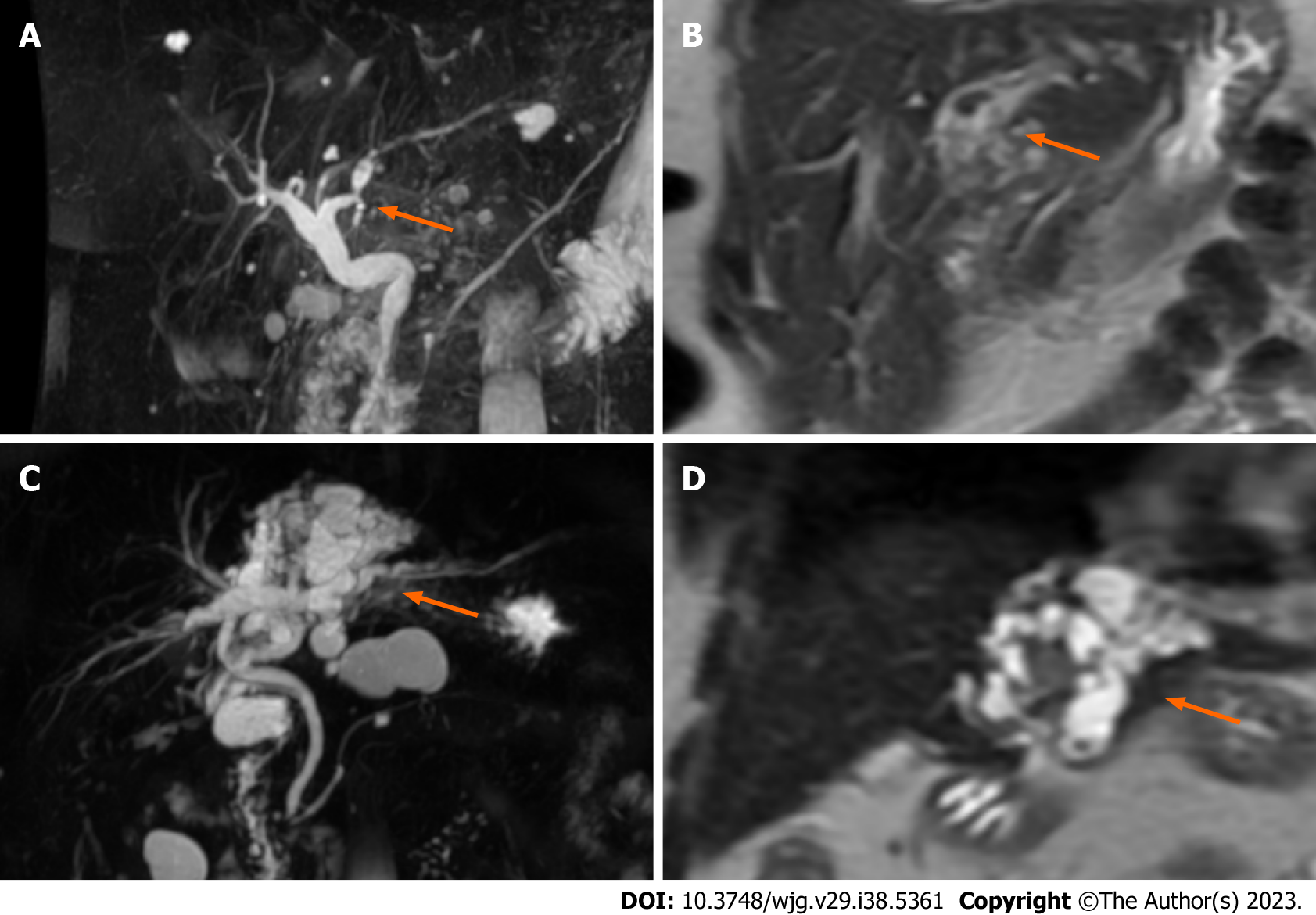
Figure 4 Magnetic resonance imaging of intraductal papillary neoplasms of the bile duct.
A: Magnetic resonance cholangiopancreatography showed an intraductal lesion of the left hepatic duct with upstream bile duct dilatation; B: Diffuse dilatation of the intrahepatic left lobe bile ducts with low intensity tumors (T2 weighted image, coronal section); C: Magnetic resonance cholangiopancreatography showed a cystic type intraductal papillary neoplasm of the bile duct; D: Cystic type intraductal papillary neoplasm of the bile duct (T2 weighted image, coronal section).
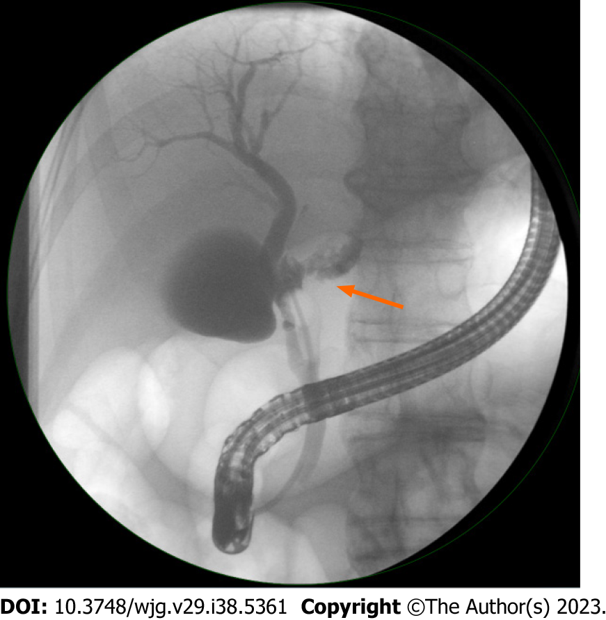
Figure 5 Endoscopic retrograde cholangiopancreatography of an intrahepatic intraductal papillary neoplasm of the bile duct showed a direct communication between the cystic lesion and the bile duct.
- Citation: Mocchegiani F, Vincenzi P, Conte G, Nicolini D, Rossi R, Cacciaguerra AB, Vivarelli M. Intraductal papillary neoplasm of the bile duct: The new frontier of biliary pathology. World J Gastroenterol 2023; 29(38): 5361-5373
- URL: https://www.wjgnet.com/1007-9327/full/v29/i38/5361.htm
- DOI: https://dx.doi.org/10.3748/wjg.v29.i38.5361


