Copyright
©The Author(s) 2023.
World J Gastroenterol. Sep 21, 2023; 29(35): 5104-5124
Published online Sep 21, 2023. doi: 10.3748/wjg.v29.i35.5104
Published online Sep 21, 2023. doi: 10.3748/wjg.v29.i35.5104
Figure 1 Effects of full-length regenerating gene 4 and nonsignal peptide regenerating gene 4 on phenotypes of DLD-1 cells.
A: DLD-1 cells were incubated with increasing doses of recombinant human regenerating gene 4 (REG4) (rhREG4) (0-500 nmol/L) for 48 h and subjected to cell proliferation assay by tetrazolium salt (MTT) assay; B: Treatment with anti-REG4 antibody produced a dose-dependent decrease in cell number of full-length (FL)-REG4-overexpressing DLD-1 cells; C: MTT assay was used to detect the proliferation of DLD-1 cells transfected with FL-REG4 or nonsignal peptide (NSP)-REG4, and treated with rhREG4 or REG4 antibody; D: Apoptosis of DLD-1 cells transfected with FL-REG4 or NSP-REG4, and treated with rhREG4 or REG4 antibody detected by flow cytometry; E: Wound healing assay was used to detect migration of DLD-1 cells transfected with FL-REG4 or NSP-REG4, and treated with rhREG4 or REG4 antibody; F: Transwell assay was used to detect migration and invasion of DLD-1 cells transfected with FL-REG4 or NSP-REG4, and treated with rhREG4 or REG4 antibody; G: Compared with DLD-1 cells, western blotting showed that transfection with FL-REG4 and treatment with rhREG4 increased expression of epidermal growth factor receptor (EGFR)-Tyr992, Tyr1068, Tyr1148, Tyr1173, Akt, p-Akt, phosphorylated phosphoinositide 3-kinase (p-PI3K), nuclear factor (NF)-κB, p-NF-KB, Bcl-2 and Bcl-X/L. REG4 antibody inhibited the effect of FL-REG4 transfection; H: Western blotting showed that compared with parental cells, DLD-1 cells transfected with NSP-REG4 had no change in expression of EGFR-Tyr992, Tyr1068, Tyr1148, Tyr1173, AKT, p-AKT, p-PI3K, NF-KB, p-NF-KB, BCL-2, or BCL-XL. aP < 0.001; bP < 0.01; dNo significance. Ab: Anti-REG4 antibody; REG4: Regenerating gene 4; rhREG4: Recombinant human regenerating gene 4; FL: Full-length; NSP: Nonsignal peptide; EGFR: Epidermal growth factor receptor; p-PI3K: Phosphorylated phosphoinositide 3-kinase; NF: Nuclear factor.
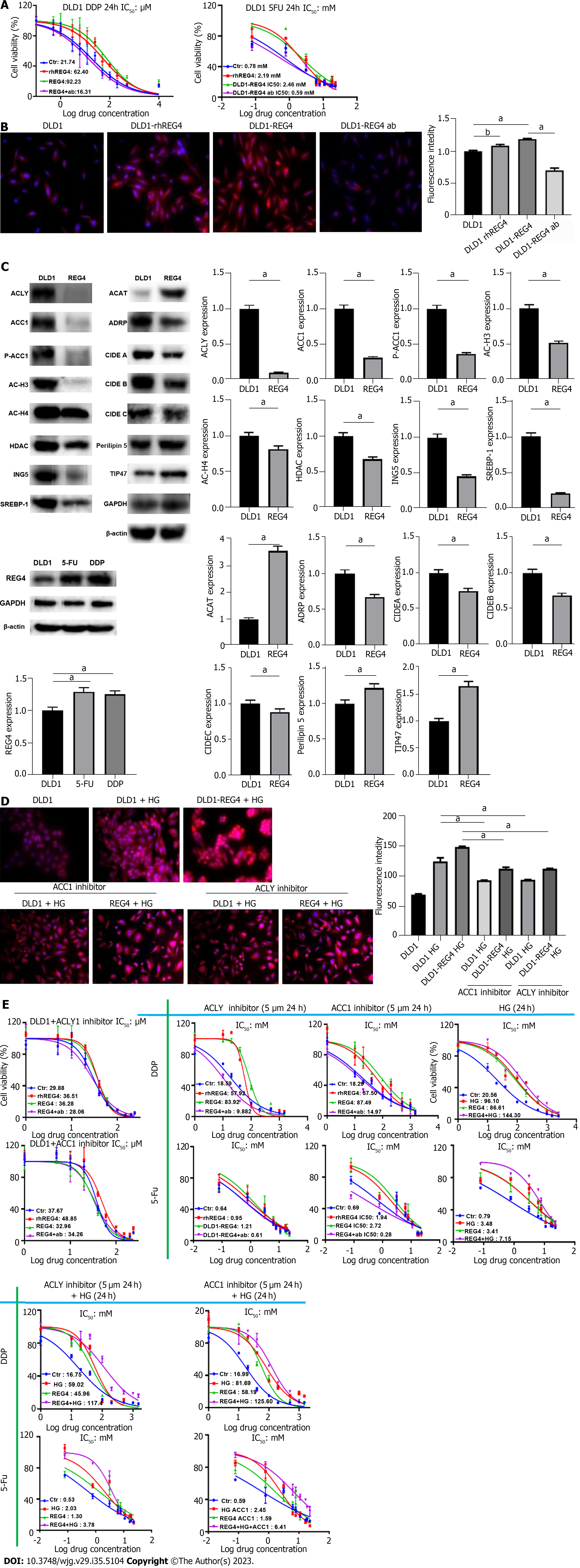
Figure 2 Effects of regenerating gene 4 on chemoresistance and droplet formation of DLD-1 cells.
A: After treatment with cisplatin (DDP) or 5-fluorouracil (5-FU), the viability was measured in DLD-1 cells, DLD-1 cells treated with recombinant human regenerating gene 4 (REG4) (rhREG4), DLD-1 cells transfected with full-length (FL)-REG4 plasmid, and DLD-1 cells treated with anti-REG4 antibody; B: The lipid droplet level was measured in DLD-1 cells, DLD-1 cells treated with rhREG4, DLD-1 cells transfected with FL-REG4 plasmid, and DLD-1 cells treated with anti-REG4 antibody; C: REG4 expression was detected in DLD-1, chemoresistant DLD-1 cells to DDP and 5-FU by western blot, proteins related to de novo synthesis or assembly pathway of lipid droplets were also detected in DLD-1 cells and DLD-1 cells transfected with FL-REG4; D: Nile red staining was used to detect the level of lipid droplets in DLD-1 cells and DLD-1 cells transfected with FL-REG4 after treatment with high glucose (HG), acetyl-CoA carboxylase 1 (ACC1) inhibitor and ATP-citrate lyase (ACLY) inhibitor; E: Tetrazolium salt assay was used to detect half maximal inhibitory concentration of DLD-1 cells for 5-FU and DDP when treated with HG, ACC1 and ACLY inhibitors, alone or in combination with the above conditions. aP < 0.001; bP < 0.01. Ab: Anti-REG4 antibody; REG4: Regenerating gene 4; rhREG4: Recombinant human regenerating gene 4; ACC1: Acetyl-CoA carboxylase 1; ACCY: ATP-citrate lyase; AC-H3: Acetyl-acetyl-histone 3; H4: Histone 4; ACC1: Acetyl-CoA carboxylase 1; HDAC: Si histone deacetylase; ING5: Inhibitor of growth protein 5; SREBP1: Sterol-regulatory element binding protein 1; ACAT: A-cholesterol acyltransferase; ADRP: Adipocyte differentiation-related protein; CIDE: Cell-death-inducing DFF45-like effector; TIP: Tail-interacting protein; DDP: Cisplatin; 5-FU: 5-fluorouracil; HG: High glucose.
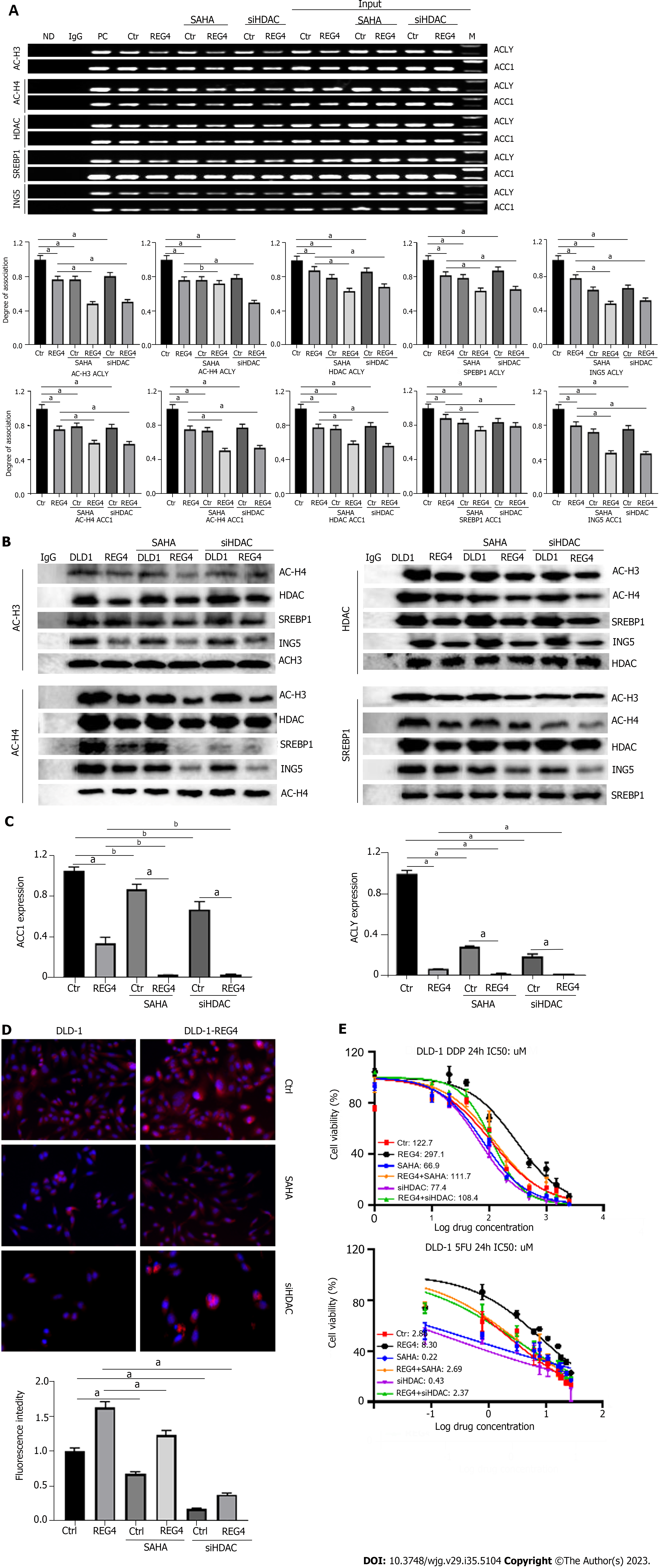
Figure 3 Full-length regenerating gene 4 weakened the transcription of acetyl-CoA carboxylase 1 and ATP-citrate lyase.
A: DLD-1 cells and full-length-regenerating gene 4 (FL-REG4) transfectants were treated with suberoylanilide hydroxamic acid (SAHA) or small interfering RNA against histone deacetylase (siHDAC), and analyzed by chromatin immunoprecipitation; B: DLD-1 cells and FL-REG4 transfectants were treated with SAHA, or siHDAC, and analyzed by co-immunoprecipitation assay using anti-anti-acetyl (AC)-acetyl-histone 3-AC-histone 4, anti-HDAC, anti-sterol-regulatory element binding protein 1 or anti-inhibitor of growth protein 5 antibody; C: DLD-1 cells and FL-REG4 transfectants were treated with SAHA or siHDAC, and analyzed by quantitative reverse transcription polymerase chain reaction; D: DLD-1 cells and FL-REG4 transfectants were treated with SAHA or siHDAC, and analyzed by Nile red staining; E: DLD-1 cells and FL-REG4 transfectants were treated with SAHA or siHDAC, and analyzed by half maximal inhibitory concentration assay of 5-fluorouracil and cisplatin. aP < 0.001; bP < 0.01. REG4: Regenerating gene 4; SAHA: Suberoylanilide hydroxamic acid; siHDAC: Small interfering RNA against histone deacetylase; IgG: Immunoglobulin G; ND: No DNA; ACC1: Acetyl-CoA carboxylase 1; ACCY: ATP-citrate lyase; AC-H3: Acetyl-acetyl-histone 3; H4: Histone 4; SREBP1: Sterol-regulatory element binding protein 1; ING5: Inhibitor of growth protein 5; Ctr: Control DLD-1 cells; PC: Positive control.
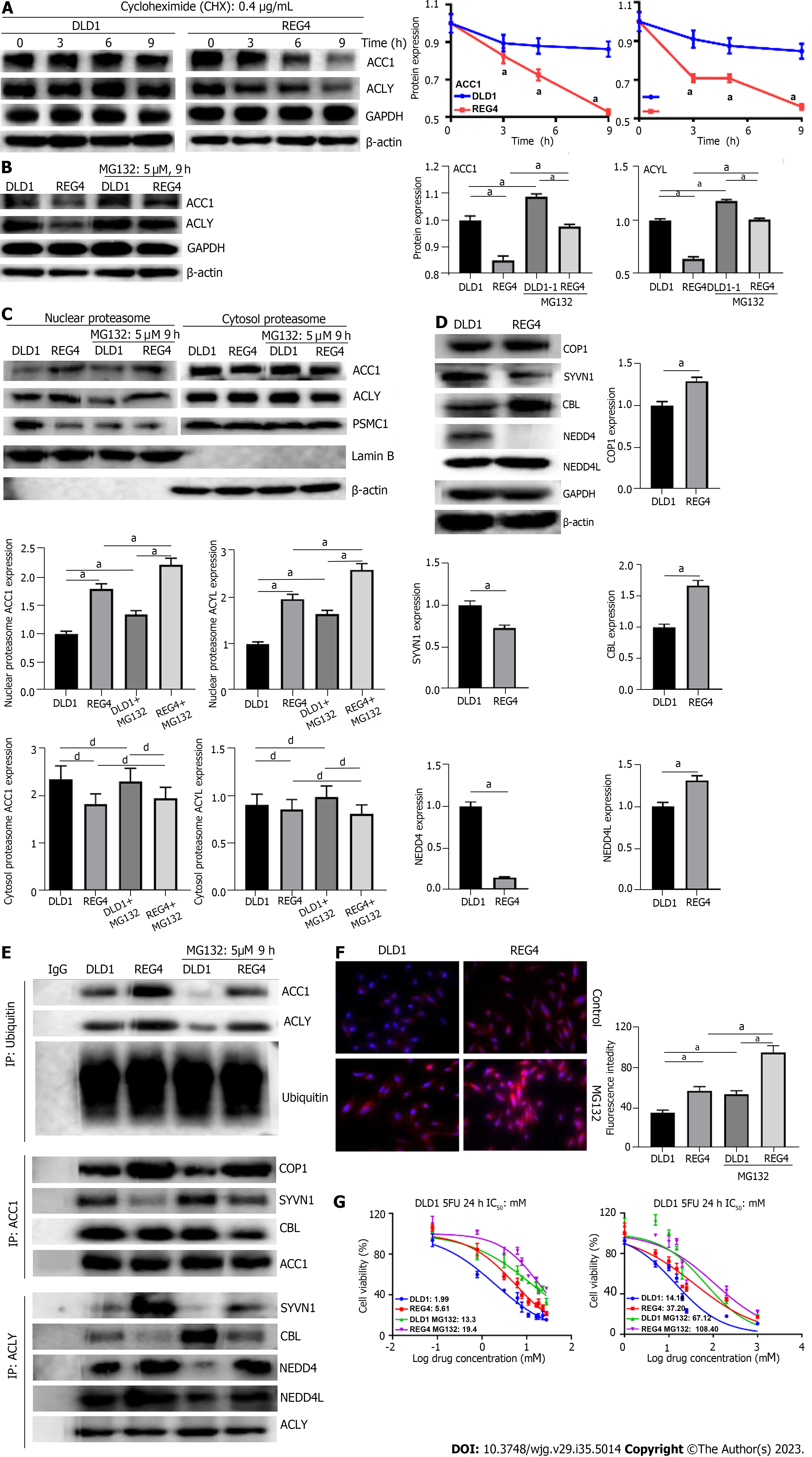
Figure 4 Full-length regenerating gene 4 destablized acetyl-CoA carboxylase 1 and ATP-citrate lyase proteins via proteasomal degradation.
A: DLD-1 cells and full-length-regenerating gene 4 (FL-REG4) transfectants were treated with cycloheximide (0.4 μg/mL), followed by western blotting to detect acetyl-CoA carboxylase 1 (ACC1) and ATP-citrate lyase (ACLY) expression; B: DLD-1 cells and FL-REG4 transfectants were treated with MG132 (5 μM, 9 h), followed by western blotting to detect ACC1 and ACLY expression; C: DLD-1 cells, FL-REG4 transfectants, and MG132-treated DLD-1 cells and FL-REG4 transfectants were subjected to proteasomal extract and western blotting to detect ACC1 and ACLY expression; D: Ubiquitin transferases (COP1 E3 ubiquitin ligase, synoviolin 1, Cbl proto-oncogene, NEDD4 like E3 ubiquitin protein ligase were detected by western blotting in DLD-1 cells and FL-REG4 transfectants; E: After co- immunoprecipitation, western blotting was performed in DLD-1 cells and FL-REG4 transfectants, with or without MG132 treatment; F: Nile red staining of DLD-1 cells, FL-REG4 transfectants, and MG132-treated DLD-1 cells and FL-REG4 transfectants; G: Tetrazolium salt assay was performed on DLD-1 cells, FL-REG4 transfectants and MG132- treated DLD-1 cells and FL-REG4 transfectants to detect half maximal inhibitory concentration of 5-fluorouracil and cisplatin. aP < 0.001; dNo significance. REG4: Regenerating gene 4; ACC1: Acetyl-CoA carboxylase 1; ACCY: ATP-citrate lyase; COP1: COP1 E3 ubiquitin ligase; SYVN1: Synoviolin 1; CBL: Cbl proto-oncogene; NEDD4L: NEDD4 like E3 ubiquitin protein ligase; IgG: Immunoglobulin G.
- Citation: Zhang CY, Zhang R, Zhang L, Wang ZM, Sun HZ, Cui ZG, Zheng HC. Regenerating gene 4 promotes chemoresistance of colorectal cancer by affecting lipid droplet synthesis and assembly. World J Gastroenterol 2023; 29(35): 5104-5124
- URL: https://www.wjgnet.com/1007-9327/full/v29/i35/5104.htm
- DOI: https://dx.doi.org/10.3748/wjg.v29.i35.5104


