Copyright
©The Author(s) 2023.
World J Gastroenterol. Sep 7, 2023; 29(33): 5014-5019
Published online Sep 7, 2023. doi: 10.3748/wjg.v29.i33.5014
Published online Sep 7, 2023. doi: 10.3748/wjg.v29.i33.5014
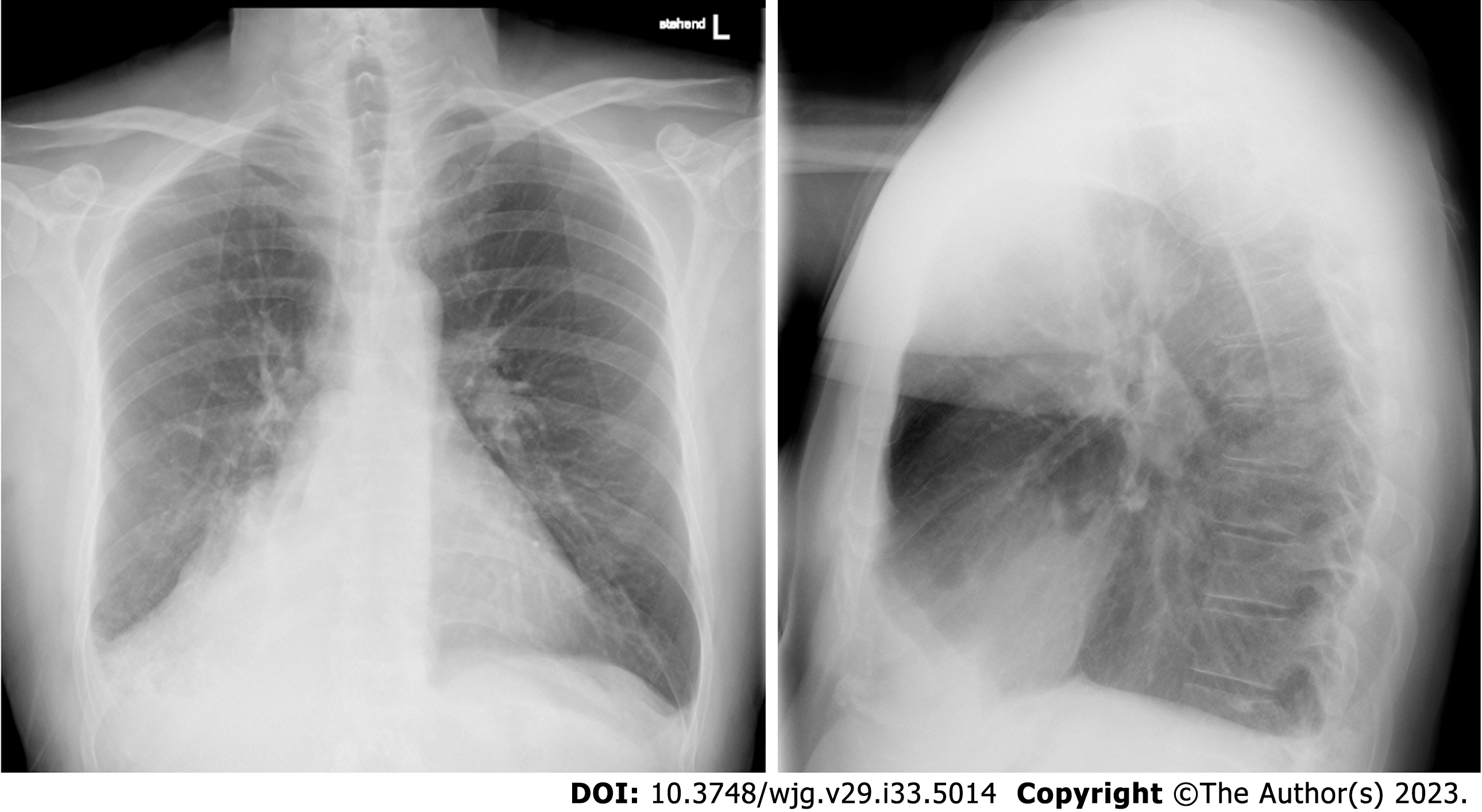
Figure 1
The initial chest roentgenogram showing an unclear visualization of the right hilar shadow suggestive of right lower lobe collapse with minimal right-sided pleural effusion.
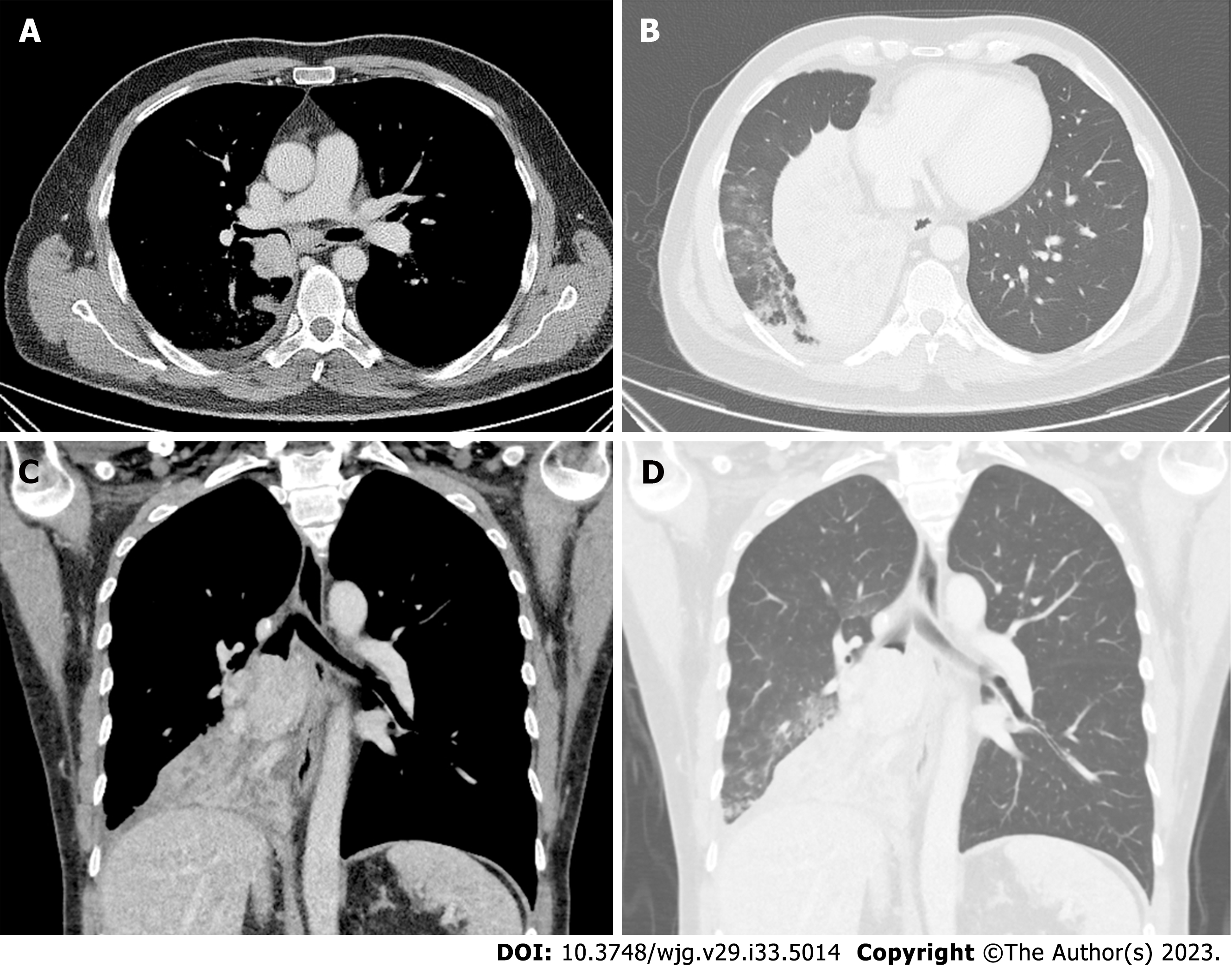
Figure 2 Preoperative computed tomography scan.
A and B: Mediastinal window during the phase of contrast shows a well-defined 43 mm × 56 mm round bordered intraluminal growth in the right bronchus intermedius occluding the airway; C and D: Lung window shows near complete collapse of the right middle lobe with no aeration post obstruction.
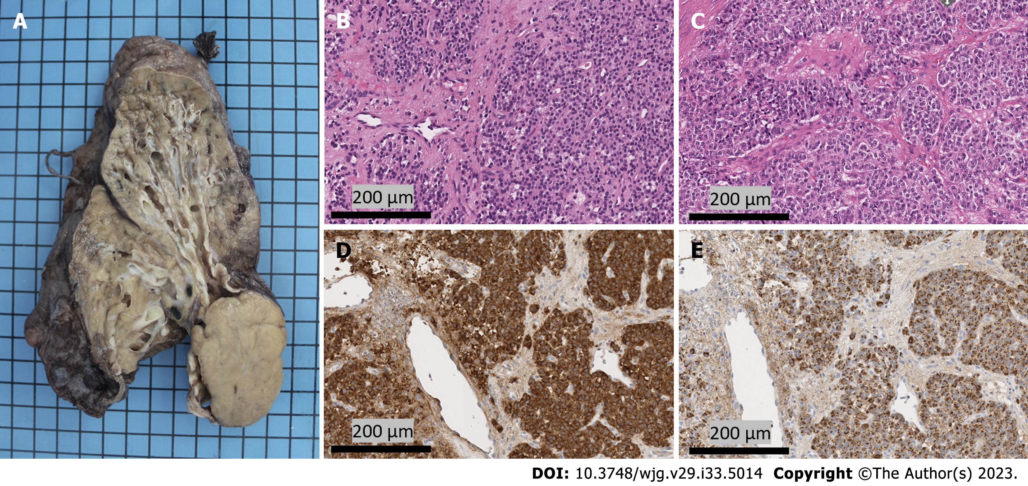
Figure 3 Surgical specimen.
A: Macroscopic appearance of a solid, well-circumscribed intraluminal tumoral lesion in the right main stem bronchus; B and C: Representative images of tumor cells revealing round nuclei, granular chromatin and large eosinophilic cytoplasm (H&E); D: Immunohistochemistry showing positivity to chromogranin (Scale 1:100); E: The tumor demonstrates high synaptophysin expression.
- Citation: Reyes CM, Klein H, Stögbauer F, Einwächter H, Boxberg M, Schirren M, Safi S, Hoffmann H. Carcinoid syndrome caused by a pulmonary carcinoid mimics intestinal manifestations of ulcerative colitis: A case report. World J Gastroenterol 2023; 29(33): 5014-5019
- URL: https://www.wjgnet.com/1007-9327/full/v29/i33/5014.htm
- DOI: https://dx.doi.org/10.3748/wjg.v29.i33.5014


