Copyright
©The Author(s) 2023.
World J Gastroenterol. Aug 28, 2023; 29(32): 4912-4919
Published online Aug 28, 2023. doi: 10.3748/wjg.v29.i32.4912
Published online Aug 28, 2023. doi: 10.3748/wjg.v29.i32.4912
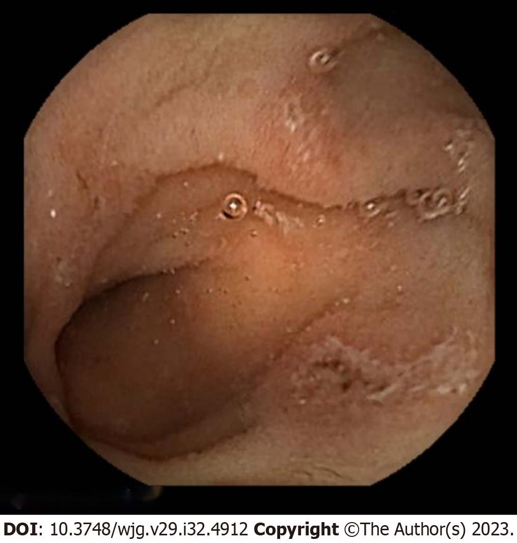
Figure 1 Colonoscopy findings at admission.
A: Distant view of colon; B: Close-up of ulcer. Multiple round punctate ulcers with a longitudinal trend from the cecum to the rectum are observed. The intervening mucosa of the ulcers is preserved, and the ulcers do not coincide with the mesenteric attachment side.
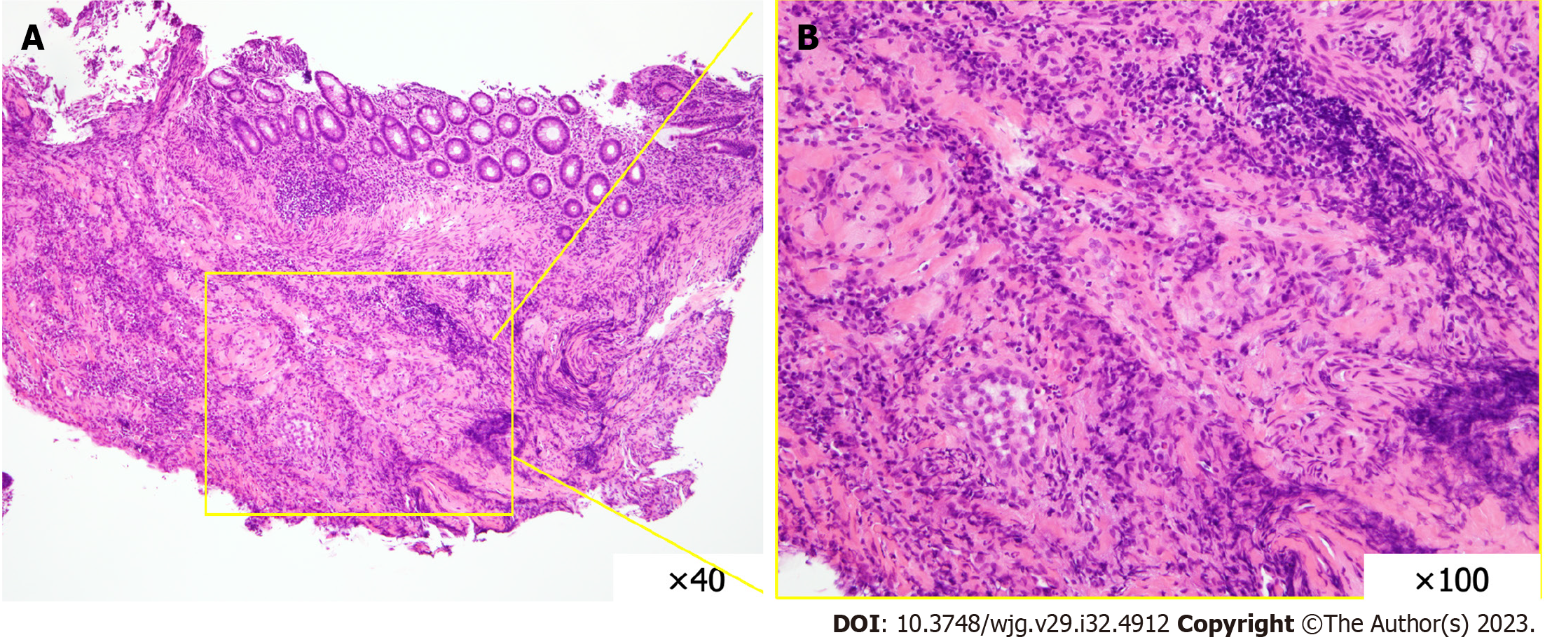
Figure 2 Pathological findings at admission.
A: Hematoxylin-eosin (HE) stained specimen at 40 × magnification; B: HE stained specimen at 100 × magnification. The mucosa is erosive and infiltrated with inflammatory cells, predominantly lymphocytes. The submucosa shows granulomatous collagen fibers and fibroblast proliferation.
Figure 3 Capsule endoscopy findings.
Scattered erosions are seen in the jejunum. The erosions were scattered on the proximal side of the jejunum; each erosion was shallow and < 1 cm in size, and hematin adhesions were visible on the surface.
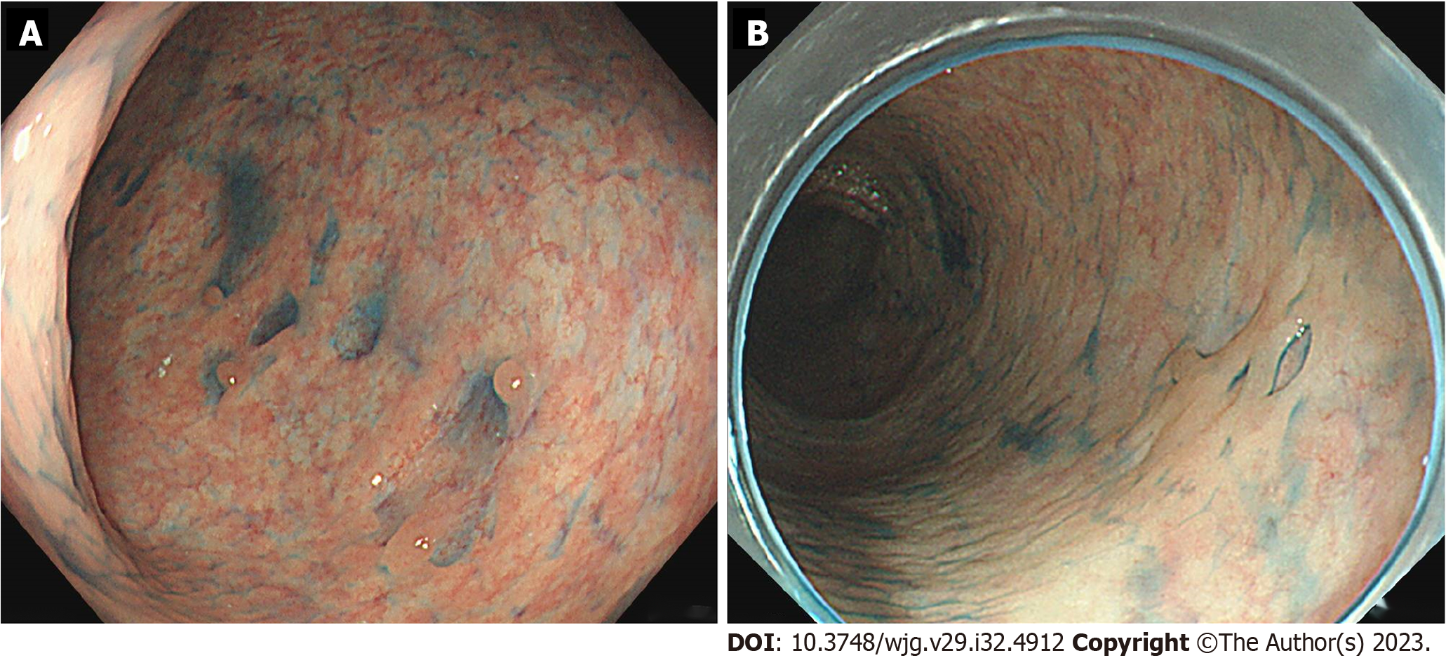
Figure 4 Colonoscopy findings after drug withdrawal.
A: At three weeks, there is shrinkage of ulcers; B: At four months, all ulcers have disappeared and scarring is observed.
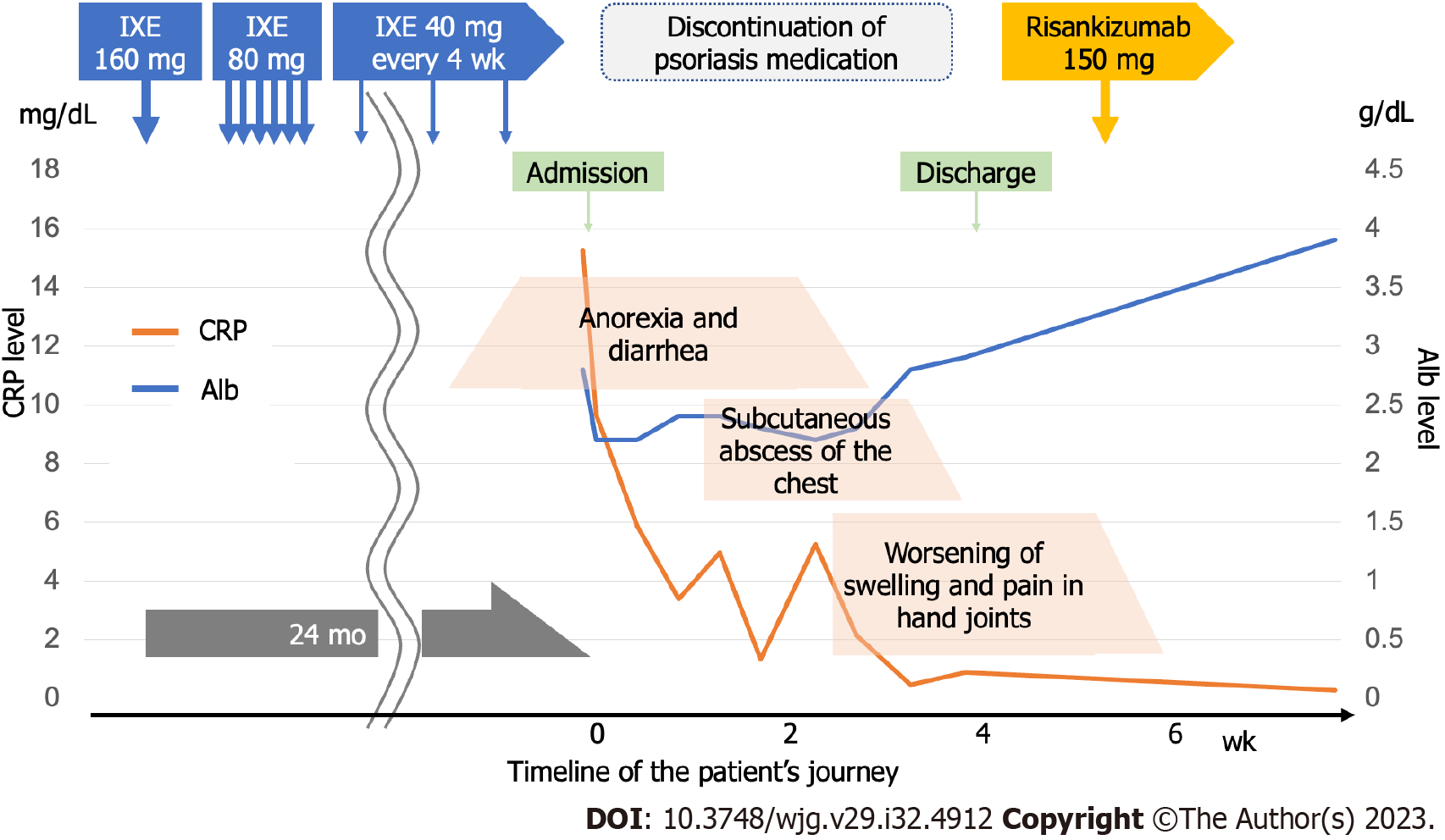
Figure 5 Albumin (Alb) and C-reactive protein (CRP) levels plotted against the timeline of the patients’ journey.
The patient visited our hospital 24 mo after the start of the ixekizumab (IXE) administration. In the graph, CRP is shown on the left vertical axis and Alb on the right vertical axis. Abdominal symptoms gradually improved with IXE withdrawal, but skin and joint symptoms tended to worsen. After 4 wk of hospitalization, the patient was discharged home, as the endoscopy showed that the ulcer had healed and the patient was able to eat adequately. After discharge, risankizumab was introduced to control skin and joint symptoms, and the patients’ condition stabilized. IXE: Ixekizumab; CRP: C-reactive protein; Alb: Albumin.
- Citation: Saito K, Yoza K, Takeda S, Shimoyama Y, Takeuchi K. Drug-induced entero-colitis due to interleukin-17 inhibitor use; capsule endoscopic findings and pathological characteristics: A case report. World J Gastroenterol 2023; 29(32): 4912-4919
- URL: https://www.wjgnet.com/1007-9327/full/v29/i32/4912.htm
- DOI: https://dx.doi.org/10.3748/wjg.v29.i32.4912


