Copyright
©The Author(s) 2023.
World J Gastroenterol. Jul 21, 2023; 29(27): 4344-4355
Published online Jul 21, 2023. doi: 10.3748/wjg.v29.i27.4344
Published online Jul 21, 2023. doi: 10.3748/wjg.v29.i27.4344
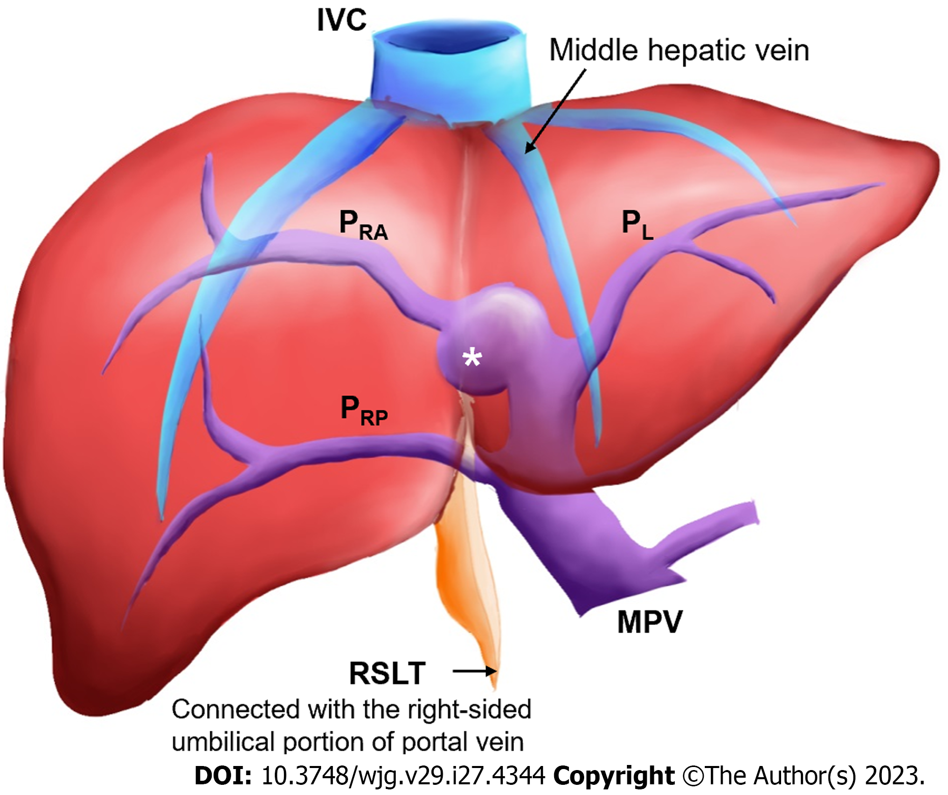
Figure 1 Coronal schematic of the most common anatomical variation in a liver with right-sided ligamentum teres.
The white asterisk indicates the umbilical portion of the portal vein. IVC: Inferior vena cava; MHV: Middle hepatic vein; PRA: Right anterior portal vein; PL: Left portal vein; PRP: Right posterior portal vein; RSLT: Right-sided ligamentum teres; MPV: Main portal vein.
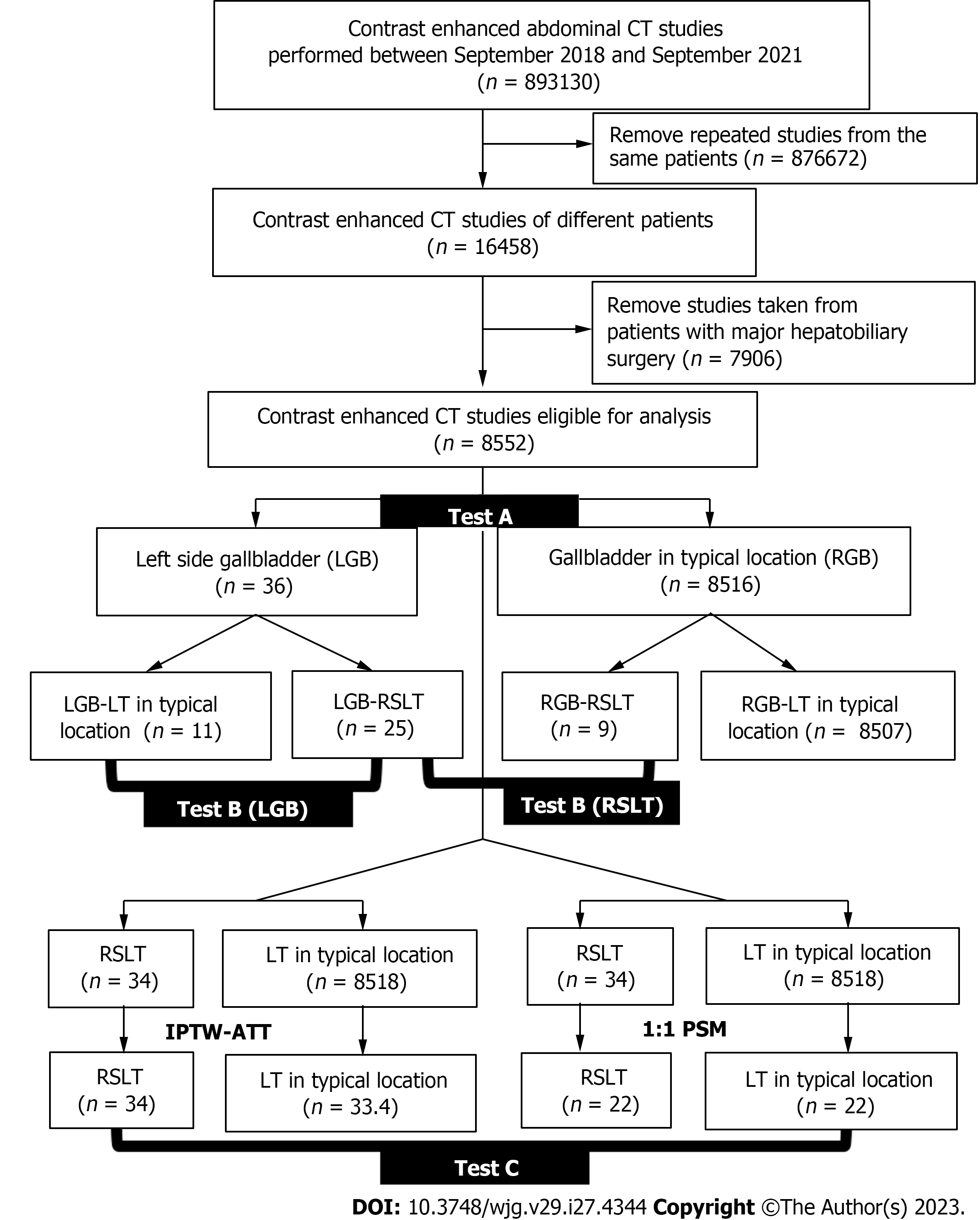
Figure 2 Flowchart of study cohort retrieval, stratification, and comparison.
RSLT: Right-sided ligamentum teres; CT: Computed tomography; LT: Ligamentum teres; PSM: Propensity score matching; LGB: Left-sided gallbladder; IPTW: Inverse probability of treatment weighting; ATT: Average treatment effect in the treated; RGB: Right-sided gallbladder.
Figure 3 Three-step method for identifying right-sided ligamentum teres, proposed by Yamashita etal[3], in axial or oblique transverse images.
A: Identify the connection of the round ligament notch to the umbilical portion of the portal vein (UP, yellow circle); B: Set an axis (dotted line) from main portal vein to the UP; C: Identify the diverging points of the dorsal branch of the right anterior portal segment (PA-D, blue arrow) and left lateral portal segment (PLL, green arrow). The diverging point of PA-D is distal to that of PLL in liver with right-sided ligamentum teres and proximal in normal liver. PA-D: Right anterior portal segment; PLL: Left lateral portal segment; UP: Umbilical portion of the portal vein; MPV: Main portal vein. Citation: Lin HY, Lee RC. Is right-sided ligamentum teres hepatis always accompanied by left-sided gallbladder? Case reports and literature review. Insights Imaging 2018; 9: 955-960. Copyright ©The Author(s) 2018. Published by Springer Nature[4]. The authors have obtained the permission for figure using (Supplementary material).
Figure 4 Intrahepatic anomalies of the portal venous system as classified by Shindoh etal[2].
A: Independent right lateral: The right lateral portal pedicle (PRL) independently originates from the main portal vein (MPV), and the right paramedian portal pedicle (PRPM) shares a trunk with the left lateral portal vein (PLL); B: Bifurcation: The MPV bifurcates into left and right portal trunks, and the PRL originates from the right portal trunk as the PRPM; C: Trifurcation: The MPV directly divides into three branches: PRL, PRPM, and PLL. PRL: Right lateral portal pedicle; PRPM: Right paramedian portal pedicle; PLL: Left lateral portal vein; RSLT: Right-sided ligamentum teres; MPV: Main portal vein. Citation: Lin HY, Lee RC. Is right-sided ligamentum teres hepatis always accompanied by left-sided gallbladder? Case reports and literature review. Insights Imaging 2018; 9: 955-960. Copyright ©The Author(s) 2018. Published by Springer Nature[4]. The authors have obtained the permission for figure using (Supplementary material).
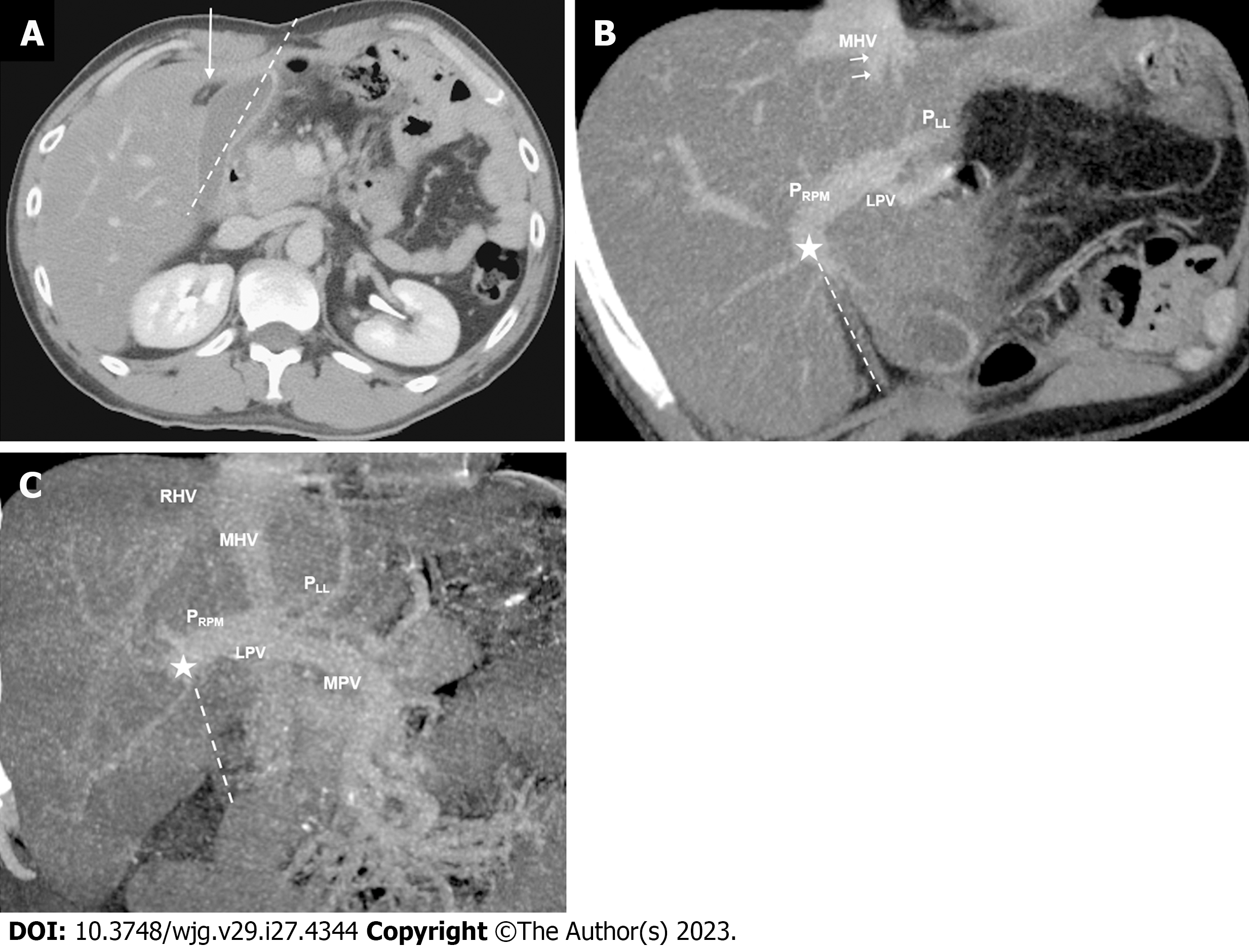
Figure 5 Contrast-enhanced computed tomography images of a 39-year-old man with right-sided ligamentum teres and left-sided gallbladder: Axial, maximum-intensity projection with oblique coronal multiplanar reformation.
A: The cholecystic axis of the gallbladder (dotted line) is located left to the umbilical fissure and right-sided ligamentum teres (RSLT, arrow); B: Right paramedian portal pedicle (PRPM) forms the right umbilical portion of the portal vein (asterisk) and joins the RSLT (dotted line). The RSLT is located right to the middle hepatic vein (MHV); C: Portal vein ramification of the Shindoh’s independent right lateral type[2]. PRPM shares a trunk with left lateral portal vein and forms the right umbilical portion of the portal vein (asterisk) before joining the RSLT (dotted line), which is located right to the MHV. PRPM: Right paramedian portal pedicle; PLL: Left lateral portal vein; MHV: Middle hepatic vein; MPV: Main portal vein; RHV: Right hepatic vein; LPV: Left portal vein.
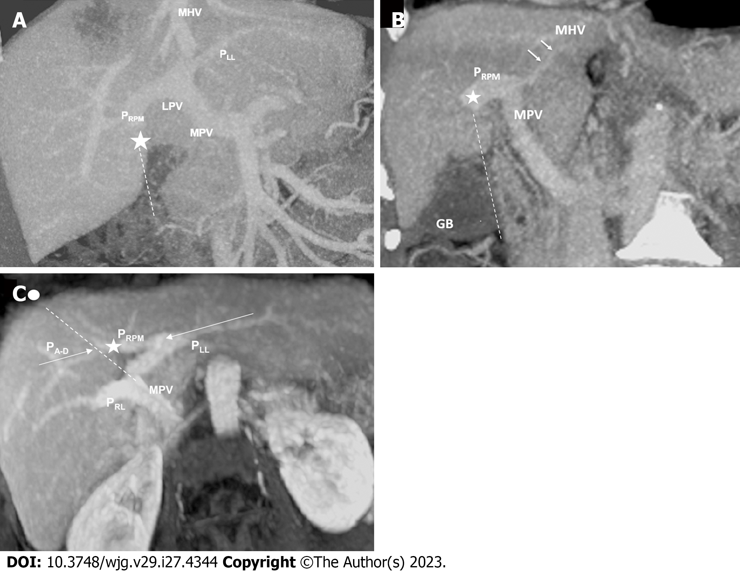
Figure 6 Contrast-enhanced computed tomography images of a 72-year-old woman with right-sided ligamentum teres and right-sided gallbladder: Maximum-intensity projection with oblique coronal multiplanar reformation, coronal maximum-intensity projection, and maximum-intensity projection with oblique axial multiplanar reformation.
A: Portal vein ramification of the Shindoh’s independent right lateral type[2]. Right paramedian portal pedicle forms the right umbilical portion of the portal vein (asterisk) and joins the right-sided ligamentum teres (RSLT, dotted line). The RSLT is located right to the middle hepatic vein (MHV); B: The cholecystic axis of the gallbladder (dotted line) is located right to the RSLT and the round ligament notch, that is, right to the MHV; C: On the axis (dotted line) along the main portal vein to the umbilical portion of the portal vein (asterisk) and the umbilical notch (circle), the diverging point of right anterior portal segment is distal to that of left lateral portal vein in this RSLT liver, consistent with the findings described by Yamashita et al[3]. PRPM: Right paramedian portal pedicle; PLL: Left lateral portal vein; PRL: Right lateral portal pedicle; PA-D: Right anterior portal segment; MHV: Middle hepatic vein; MPV: Main portal vein; GB: Gallbladder; LPV: Left portal vein.
- Citation: Lin HY, Lee RC, Chai JW, Hsu CY, Chou Y, Hwang HE, Liu CA, Chiu NC, Yen HH. Predicting portal venous anomalies by left-sided gallbladder or right-sided ligamentum teres hepatis: A large scale, propensity score-matched study. World J Gastroenterol 2023; 29(27): 4344-4355
- URL: https://www.wjgnet.com/1007-9327/full/v29/i27/4344.htm
- DOI: https://dx.doi.org/10.3748/wjg.v29.i27.4344


