Copyright
©The Author(s) 2023.
World J Gastroenterol. May 28, 2023; 29(20): 3066-3083
Published online May 28, 2023. doi: 10.3748/wjg.v29.i20.3066
Published online May 28, 2023. doi: 10.3748/wjg.v29.i20.3066
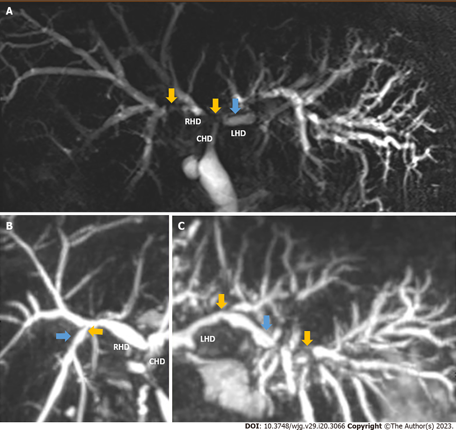
Figure 1 Magnetic resonance cholangiopancreatography reconstruction images of ischemic type biliary lesions in donated after circulatory death graft recipients.
A: Significant structuring and confluence of right and left ducts, in addition to the right posterior duct (yellow arrows). Upstream dilation on left indicated by blue arrow; B: Structuring at a second order duct on the right (yellow arrow) with upstream dilation; C: Strictures in periphery of left biliary system. Figure created with biorender.com, accessed on January 2023. LHD: Left ducts; RHD: Right ducts; CHD: Common hepatic duct.
- Citation: Durán M, Calleja R, Hann A, Clarke G, Ciria R, Nutu A, Sanabria-Mateos R, Ayllón MD, López-Cillero P, Mergental H, Briceño J, Perera MTPR. Machine perfusion and the prevention of ischemic type biliary lesions following liver transplant: What is the evidence? World J Gastroenterol 2023; 29(20): 3066-3083
- URL: https://www.wjgnet.com/1007-9327/full/v29/i20/3066.htm
- DOI: https://dx.doi.org/10.3748/wjg.v29.i20.3066


