Copyright
©The Author(s) 2023.
World J Gastroenterol. Mar 21, 2023; 29(11): 1757-1764
Published online Mar 21, 2023. doi: 10.3748/wjg.v29.i11.1757
Published online Mar 21, 2023. doi: 10.3748/wjg.v29.i11.1757
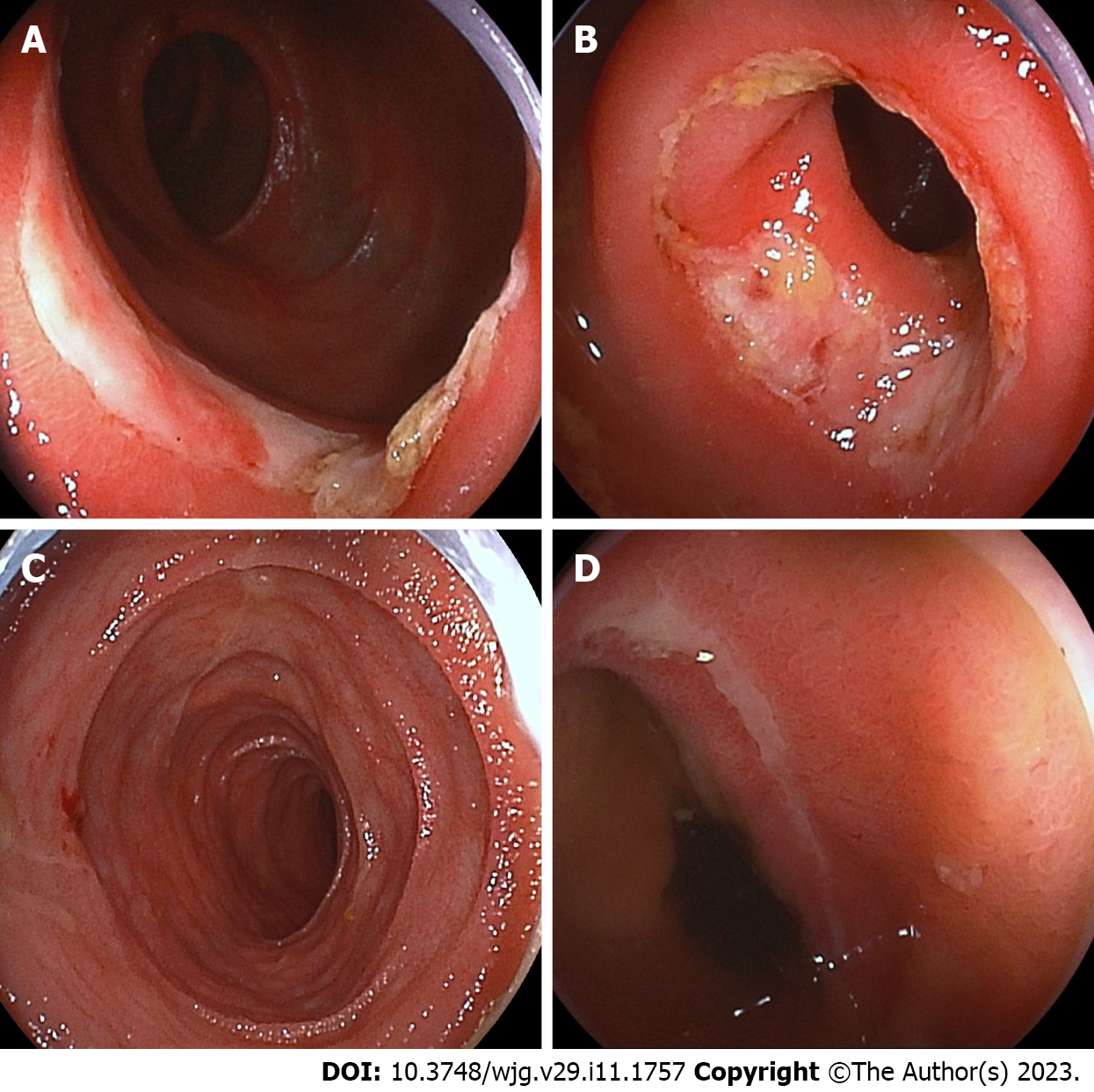
Figure 1 Findings of initial double-balloon small intestinal endoscopy.
A: Initial double-balloon enteroscopy (DBE) shows multiple oblique ulcers with discrete margins, 70-100 cm proximal to the ileal valve; B: The circular ulcers with slight constriction of the small intestinal lumen at initial DBE; C: Follow-up DBE performed 1 year later shows mucosal healing; D: The oblique ulcer and scars at 70 cm proximal to the ileal valve at follow-up DBE.
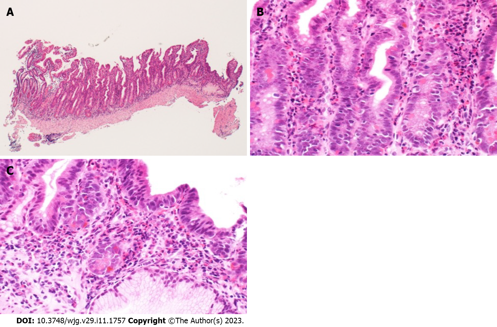
Figure 2 Histopathological findings of ileal tissue obtained from double-balloon enteroscopy.
A: Hematoxylin and eosin staining (× 100) shows mild villous atrophy; B: Hematoxylin and eosin staining (× 400) shows moderate to severe histological eosinophilic infiltration (maximum 80 eosinophils/high-power field); C: Hematoxylin and eosin staining (× 400) shows crypt destruction (cryptitis).
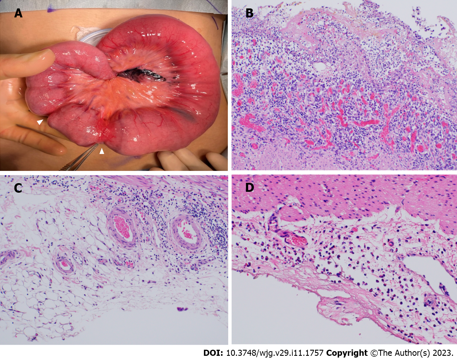
Figure 3 Macroscopic and microscopic histological findings of the resected ileum.
A: Macroscopically, strictures are observed 40 cm and 44 cm proximal to the ileocecal valve (arrowhead); B: Resected ileum shows ulcer formation and peri-ulcer mucosal damage histologically; C: No significant eosinophilic infiltration is observed transmurally; D: Resected ileum at the ileostomy closure shows marked eosinophilic infiltration (> 50/high-power field) in the subserosa.
- Citation: Kimura K, Jimbo K, Arai N, Sato M, Suzuki M, Kudo T, Yano T, Shimizu T. Eosinophilic enteritis requiring differentiation from chronic enteropathy associated with SLCO2A1 gene: A case report. World J Gastroenterol 2023; 29(11): 1757-1764
- URL: https://www.wjgnet.com/1007-9327/full/v29/i11/1757.htm
- DOI: https://dx.doi.org/10.3748/wjg.v29.i11.1757


