Copyright
©The Author(s) 2022.
World J Gastroenterol. Mar 7, 2022; 28(9): 909-917
Published online Mar 7, 2022. doi: 10.3748/wjg.v28.i9.909
Published online Mar 7, 2022. doi: 10.3748/wjg.v28.i9.909
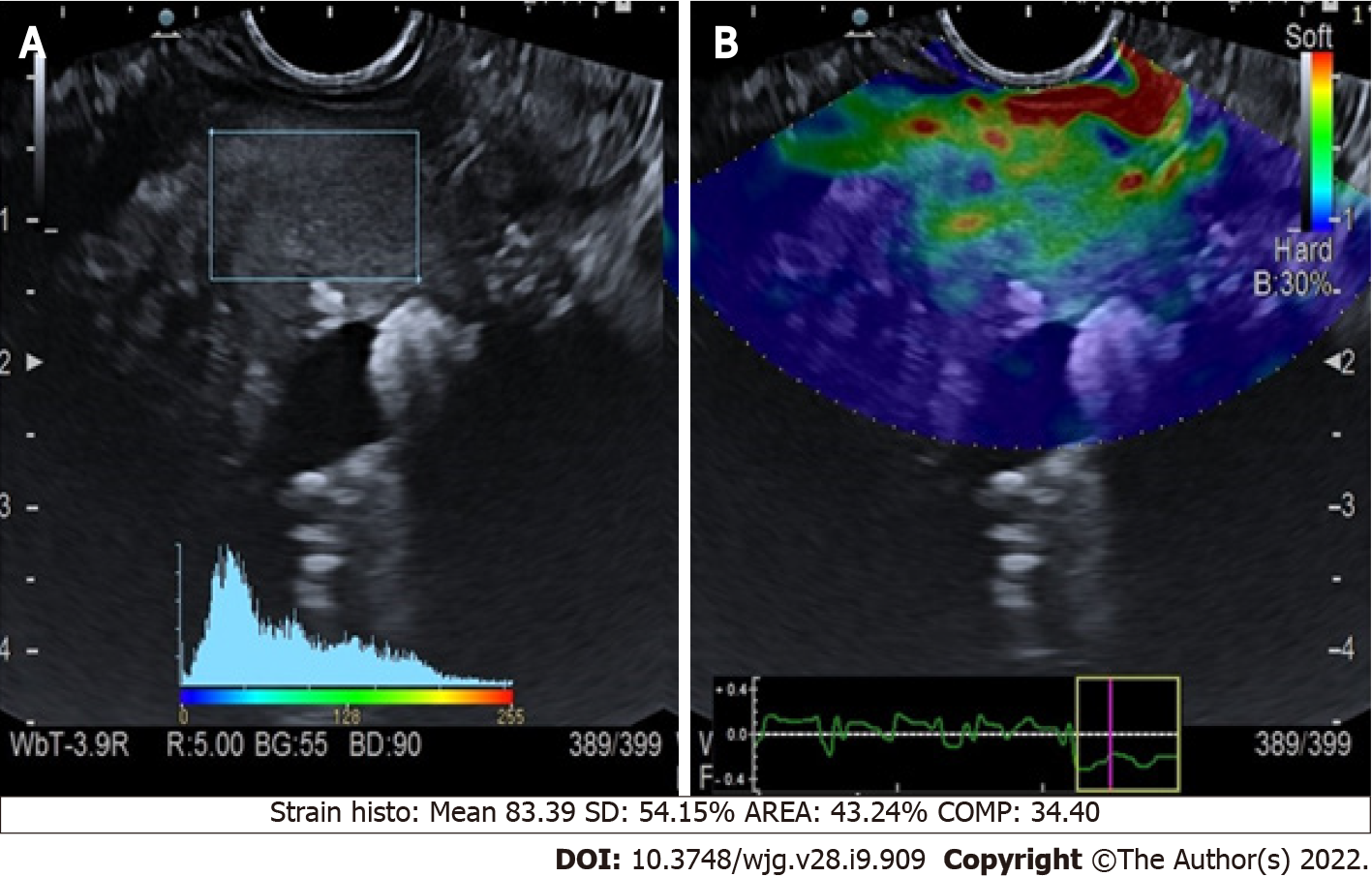
Figure 1 Quantitative endoscopic ultrasound elastography of chronic pancreatitis.
A: Endoscopic ultrasound B-mode with region of interest square; B: Elastographic image.
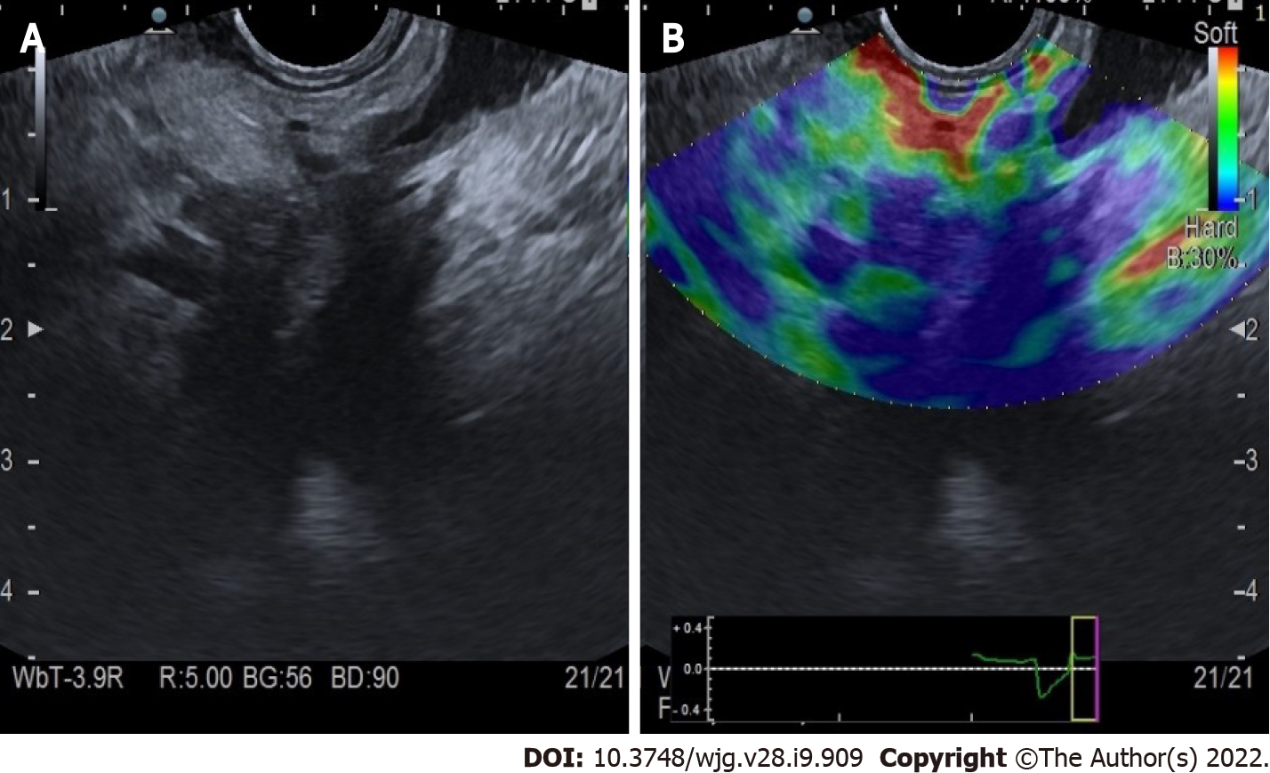
Figure 2 Qualitative endoscopic ultrasound elastography of pancreatic adenocarcinoma of the head.
A: Endoscopic ultrasound B-mode; B: Elastographic image.
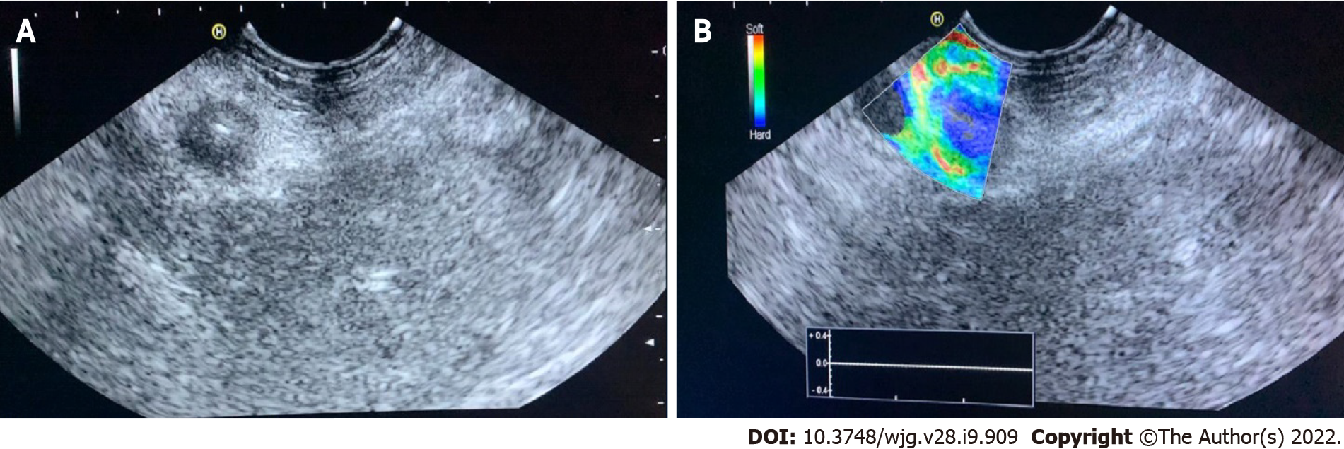
Figure 3 Qualitative endoscopic ultrasound elastography of a neuroendocrine malignant tumor of the pancreatic tail.
A: Endoscopic ultrasound B-mode; B: Elastographic image.
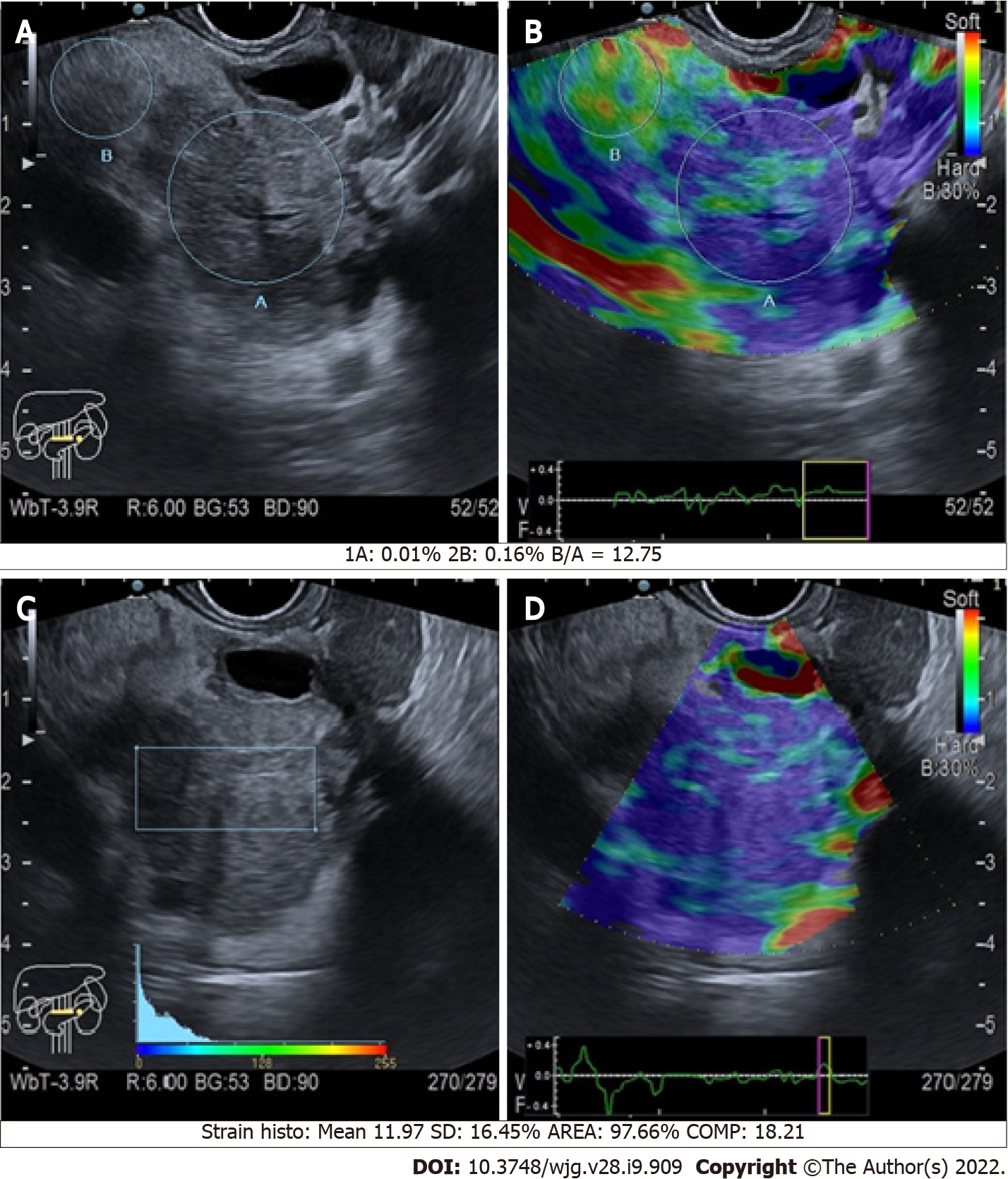
Figure 4 Quantitative endoscopic ultrasound elastography of pancreatic cancers of the body.
A and C: B-mode with region of interest circles and square; B and D: Elastographic images.
- Citation: Conti CB, Mulinacci G, Salerno R, Dinelli ME, Grassia R. Applications of endoscopic ultrasound elastography in pancreatic diseases: From literature to real life. World J Gastroenterol 2022; 28(9): 909-917
- URL: https://www.wjgnet.com/1007-9327/full/v28/i9/909.htm
- DOI: https://dx.doi.org/10.3748/wjg.v28.i9.909


