Copyright
©The Author(s) 2022.
World J Gastroenterol. Dec 21, 2022; 28(47): 6632-6661
Published online Dec 21, 2022. doi: 10.3748/wjg.v28.i47.6632
Published online Dec 21, 2022. doi: 10.3748/wjg.v28.i47.6632
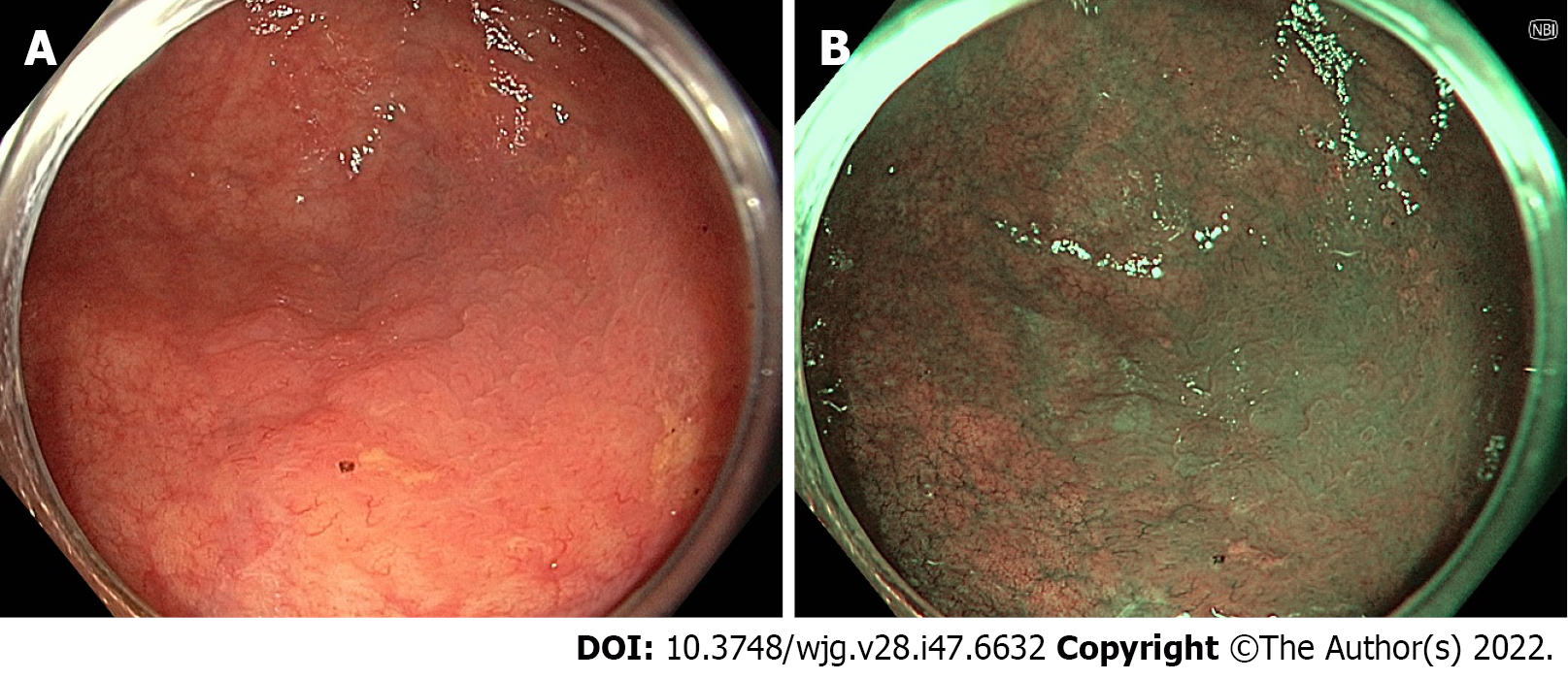
Figure 1 Sessile serrated adenoma/polyp seen on high-definition white light imaging and narrow-band imaging.
A: High-definition white light imaging; B: Narrow-band imaging.
Figure 2 Sessile serrated adenoma seen on white light imaging, linked colour imaging, and blue light imaging.
A: White light imaging; B: Linked colour imaging; C: Blue light imaging.
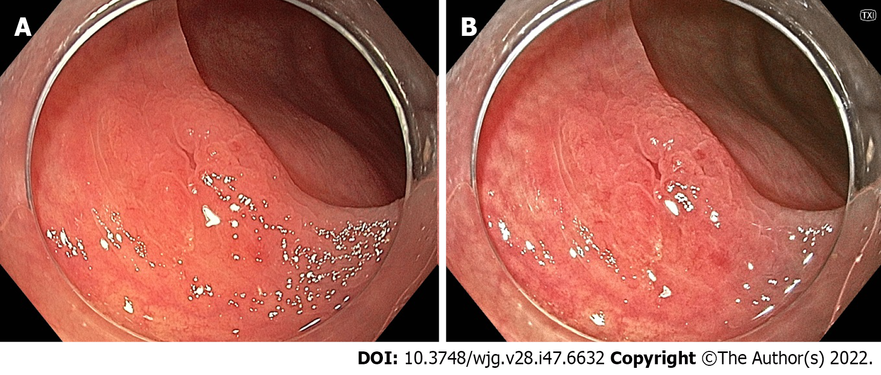
Figure 3 Sessile serrated adenoma seen on white light imaging and texture and colour enhancement imaging.
A: White light imaging; B: Texture and colour enhancement imaging.
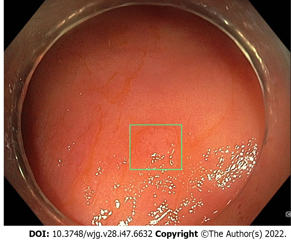
Figure 4 Computer aided detection detection system with a real-time alert seen around a flat tubular adenoma.
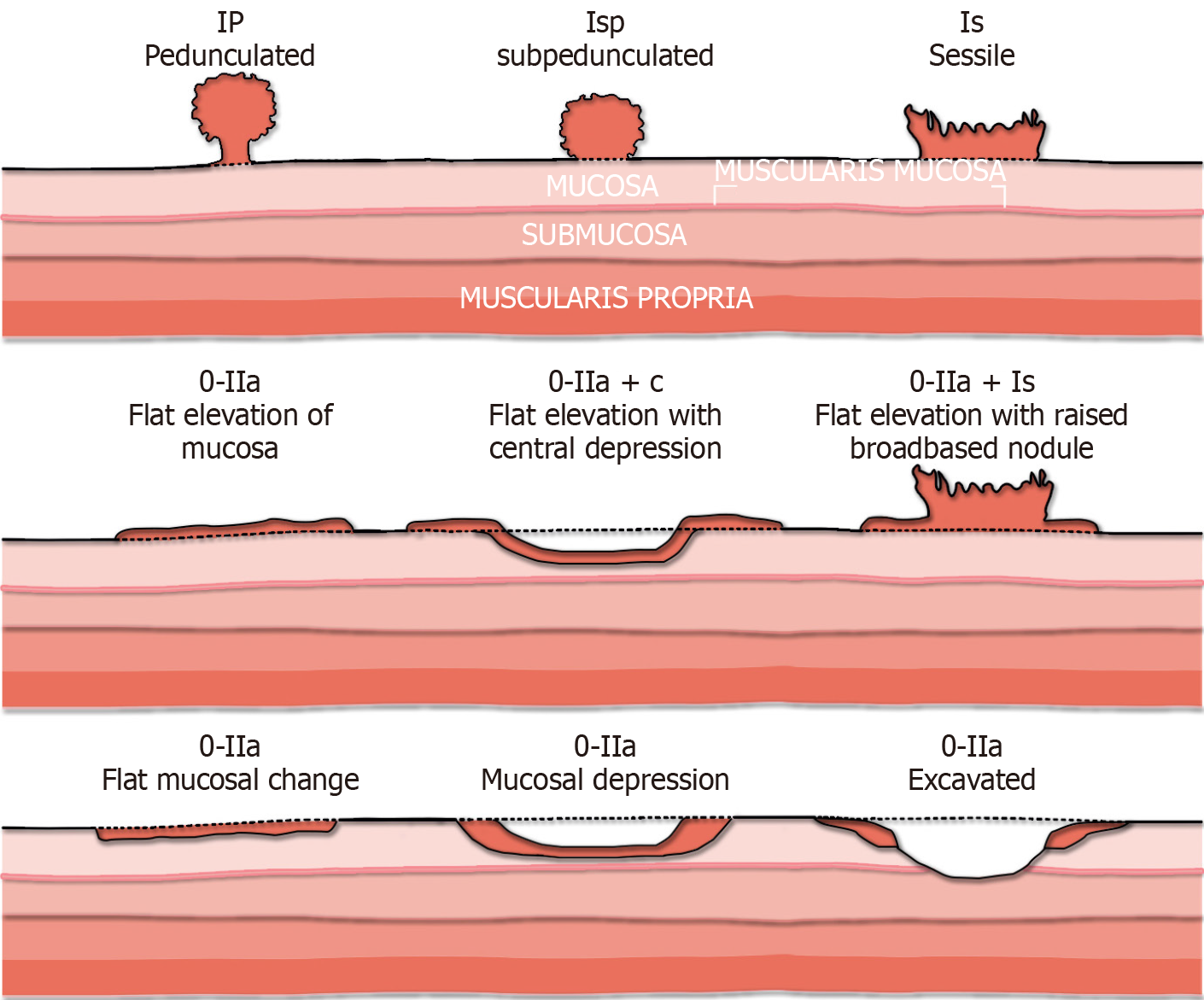
Figure 5 Paris classification.
Citation: Mathews AA, Draganov PV, Yang D. Endoscopic management of colorectal polyps: From benign to malignant polyps. World J Gastrointest Endosc 2021; 13: 356-370[141].
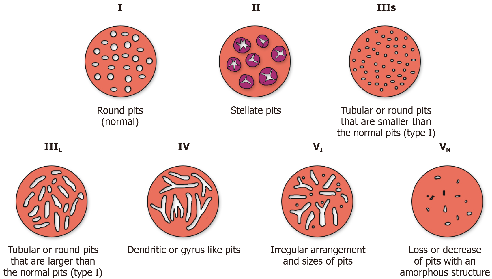
Figure 6 Kudo’s classification.
Citation: Mathews AA, Draganov PV, Yang D. Endoscopic management of colorectal polyps: From benign to malignant polyps. World J Gastrointest Endosc 2021; 13: 356-370[141].
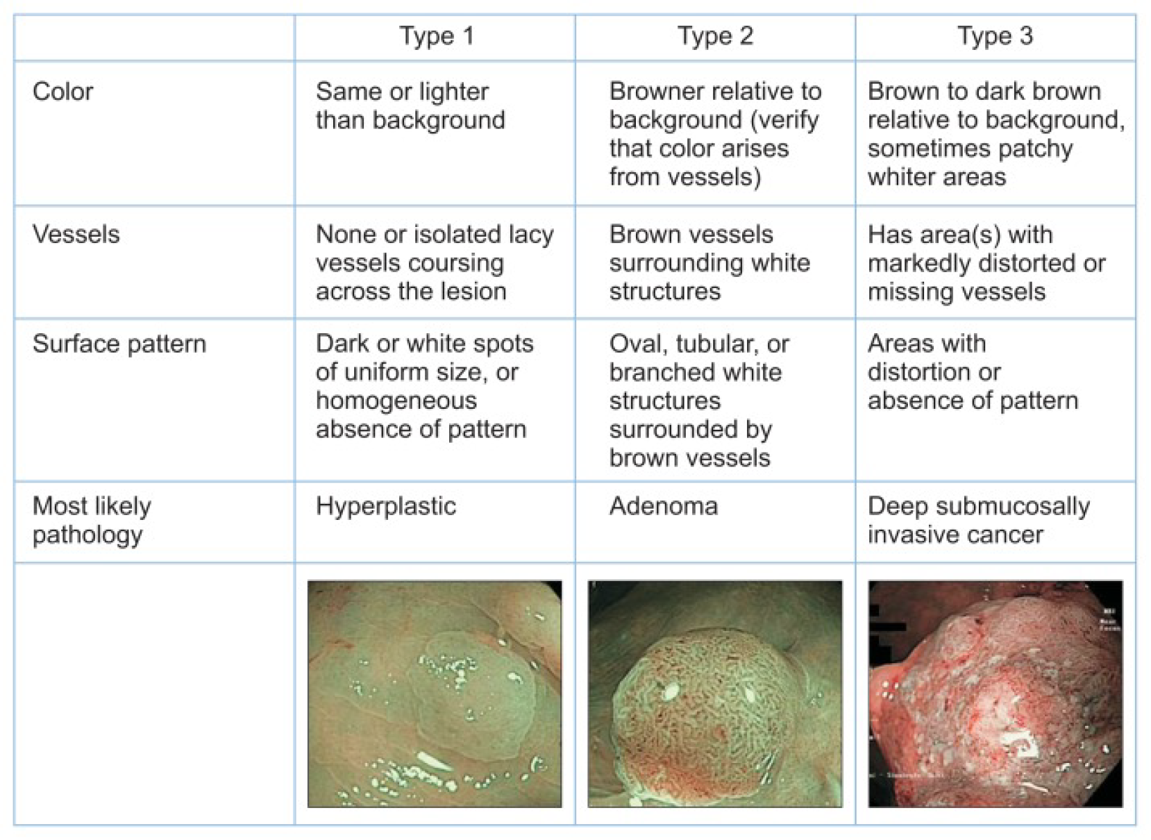
Figure 7 Narrow-band Imaging International Colorectal Endoscopic classification.
Citation: Puig I, Kaltenbach T. Optical Diagnosis for Colorectal Polyps: A Useful Technique Now or in the Future? Gut Liver 2018; 12: 385-392[150]. Copyright© The Author(s) 2018. Published by The Korean Society of Gastroenterology, the Korean Society of Gastrointestinal Endoscopy, the Korean Society of Neurogastroenterology and Motility, Korean College of Helicobacter and Upper Gastrointestinal Research, Korean Association the Study of Intestinal Diseases, the Korean Association for the Study of the Liver, Korean Pancreatobiliary Association, and Korean Society of Gastrointestinal Cancer (Supplementary material).
Figure 8 Japan Narrow-band Imaging Expert Team developed the Japan Narrow-band Imaging expert team classification.
Citation: Hirata D, Kashida H, Iwatate M, Tochio T, Teramoto A, Sano Y, Kudo M. Effective use of the Japan Narrow Band Imaging Expert Team classification based on diagnostic performance and confidence level. World J Clin Cases 2019; 7: 2658-2665[158]. Copyright© The Author(s) 2019. Published by Baishideng Publishing Group Inc (Supplementary material).
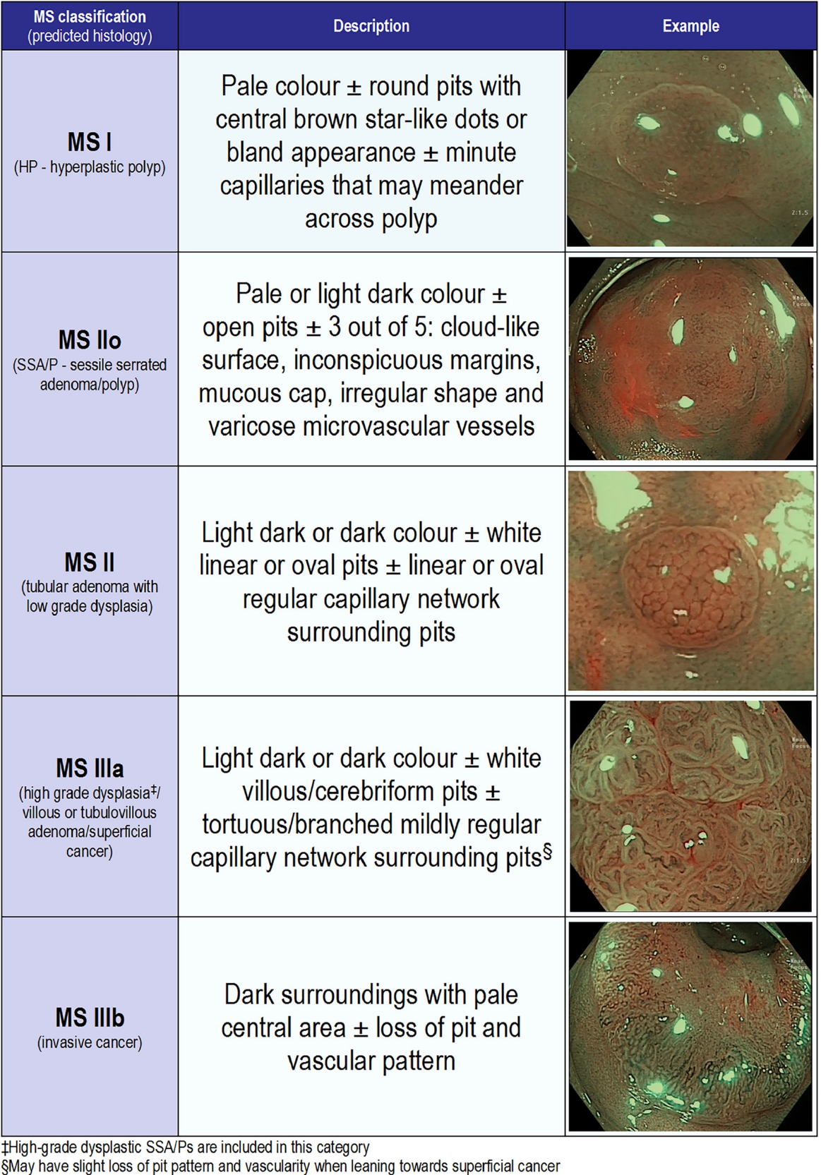
Figure 9 mSano classification demonstrating delineation between sessile serrated adenomas/polyps and hyperplastic polyps.
Citation: Zorron Cheng Tao Pu L, Yamamura T, Nakamura M, Koay DSC, Ovenden A, Edwards S, Burt AD, Hirooka Y, Fujishiro M, Singh R. Comparison of different virtual chromoendoscopy classification systems for the characterization of colorectal lesions. JGH Open 2020; 4: 818-826[168]. Copyright© The Author(s) 2019. Published by Journal of Gastroenterology and Hepatology Foundation and John Wiley & Sons Australia, Ltd (Supplementary material).
Figure 10 Large colonic laterally spreading tumour.
A: White light imaging with poor differentiation between polyp and normal tissue; B: Flat extension seen more clearly on NBI; C: Submucosal methylene blue injection prior to resection clearly delineating the margins of the flat spreading component.
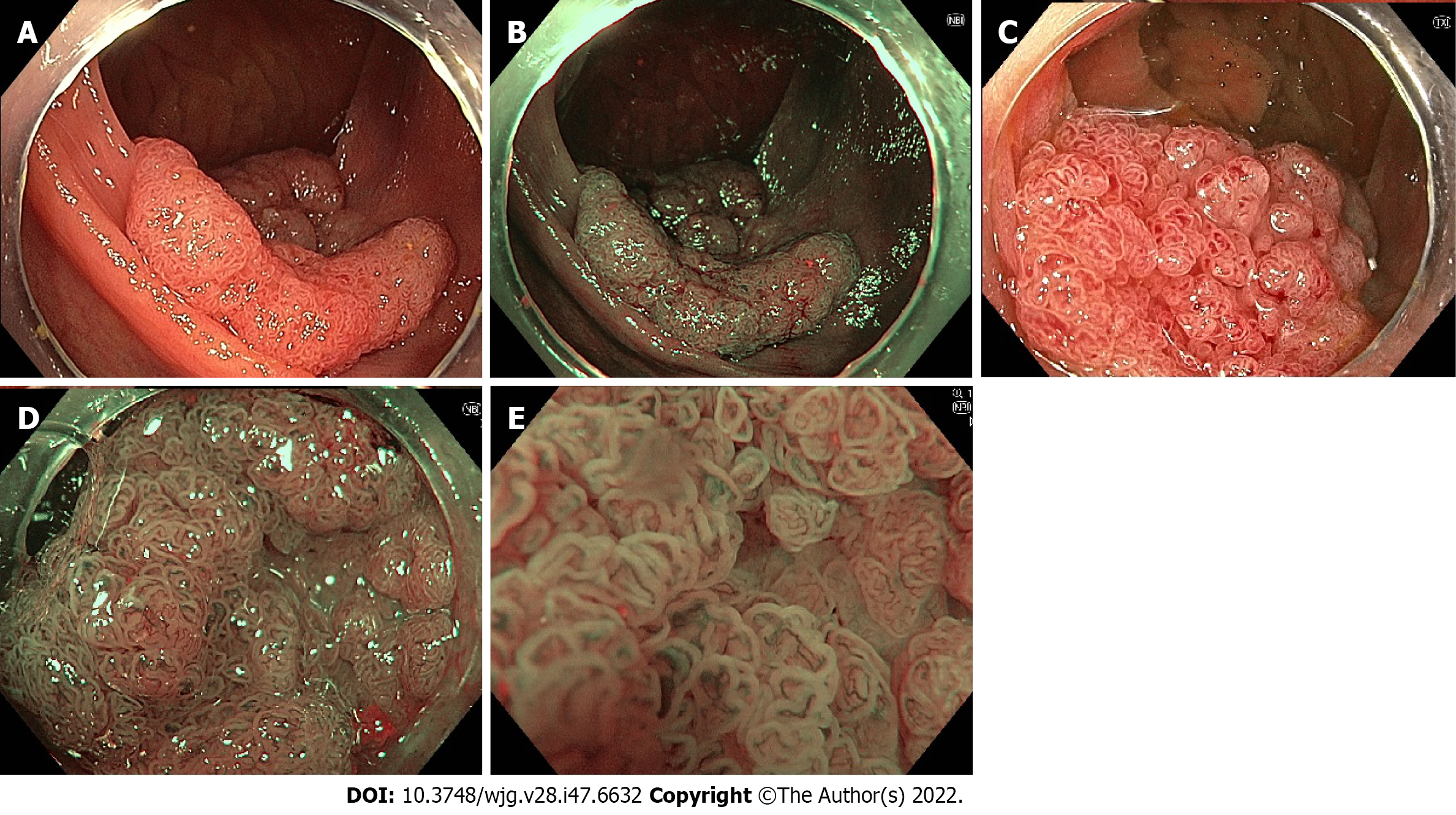
Figure 11 Laterally spreading tubulo-villous adenoma with high-grade dysplasia.
A: White light imaging; B: Narrow-band imaging (NBI); C: Texture and colour enhancement imaging; D: NBI with magnification; E: NBI with high-magnification using the underwater technique.
Figure 12 Tubulo-villous adenoma.
A: White light imaging; B: Blue light imaging; C: Linked colour imaging.
- Citation: Young EJ, Rajandran A, Philpott HL, Sathananthan D, Hoile SF, Singh R. Mucosal imaging in colon polyps: New advances and what the future may hold. World J Gastroenterol 2022; 28(47): 6632-6661
- URL: https://www.wjgnet.com/1007-9327/full/v28/i47/6632.htm
- DOI: https://dx.doi.org/10.3748/wjg.v28.i47.6632


