Copyright
©The Author(s) 2022.
World J Gastroenterol. Dec 14, 2022; 28(46): 6433-6477
Published online Dec 14, 2022. doi: 10.3748/wjg.v28.i46.6433
Published online Dec 14, 2022. doi: 10.3748/wjg.v28.i46.6433
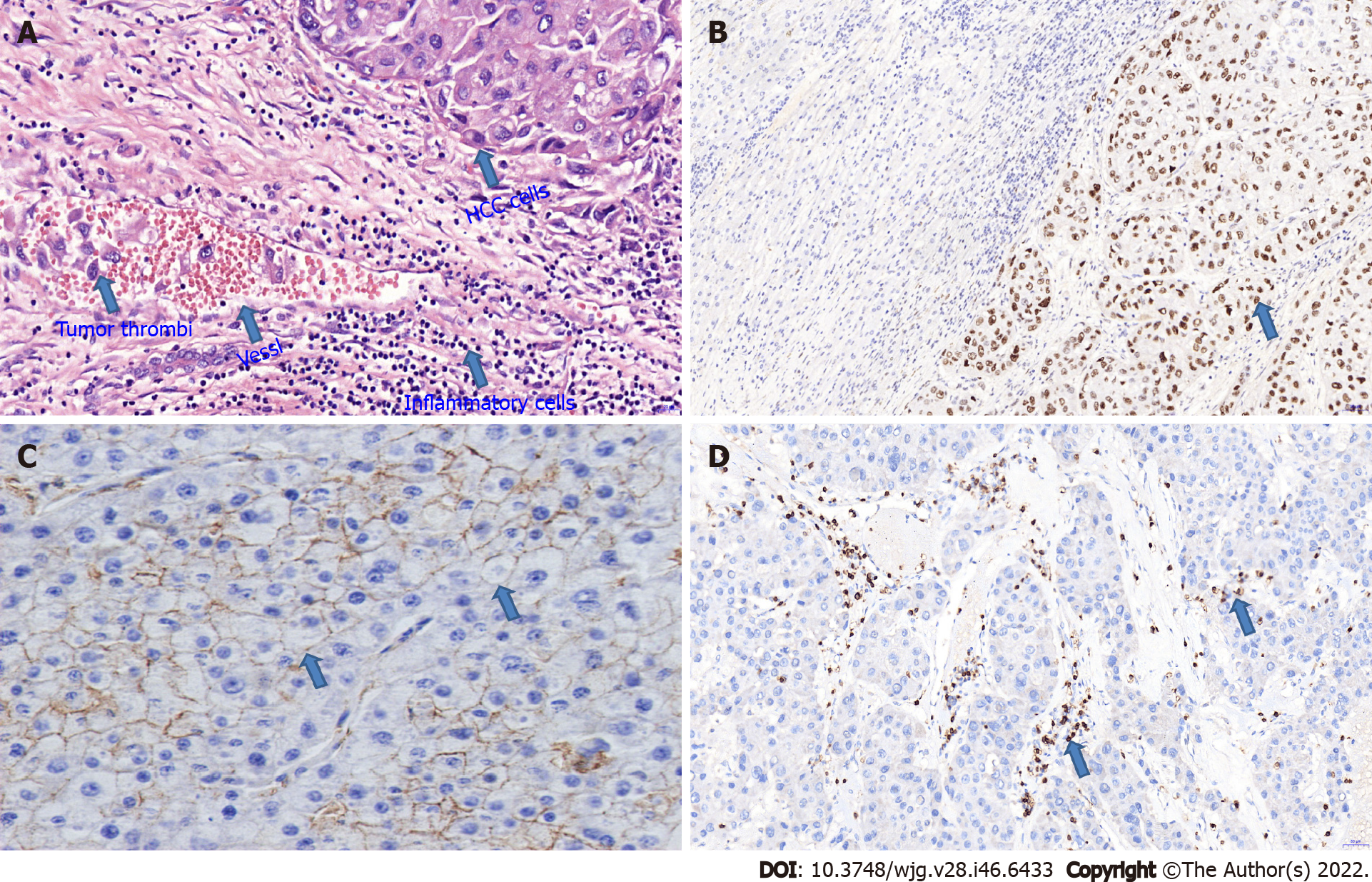
Figure 1 Microscopic image.
A: Microscopic image of intrahepatic hematogenous metastasis. The presence of tumor thrombi in the vessels indicates that the Hepatocellular carcinoma (HCC) cells have invaded the surrounding blood vessels and lymphatic vessels, which is a high-risk factor for HCC recurrence and metastasis (arrow, hematoxylin and eosin × 400); B: Microscopic image of mutant p53 expression in HCC cells. TP53 is highly expressed in HCC tissues (Right), but not in benign liver tissues (Left) (arrow, immunohistochemical staining × 200); C: Microscopic image of KAI1 expression in HCC cells. KAI1 expression is down-regulated or absent in HCC cells (arrow, immunohistochemical staining × 200); D: Microscopic image of CD3+ T cells in HCC tissues and stromal tissues. CD3+ T cells are densely distributed in tumor stroma, while a single CD3+ T cell is occasionally distributed among HCC cells (arrow, immunohistochemical staining × 200). HCC: Hepatocellular carcinoma.
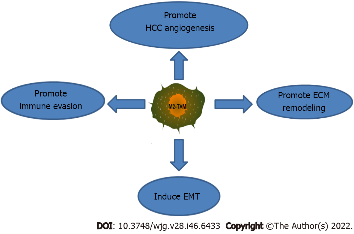
Figure 2 Schematic diagram of molecular mechanisms by which M2-type tumor associated macrophages promote hepatocellular carcinoma metastasis and recurrence.
HCC: Hepatocellular carcinoma; M2-TAMs: M2-type tumor associated macrophages; ECM: Extracellular matrix; EMT: Epithelial mesenchymal transition.
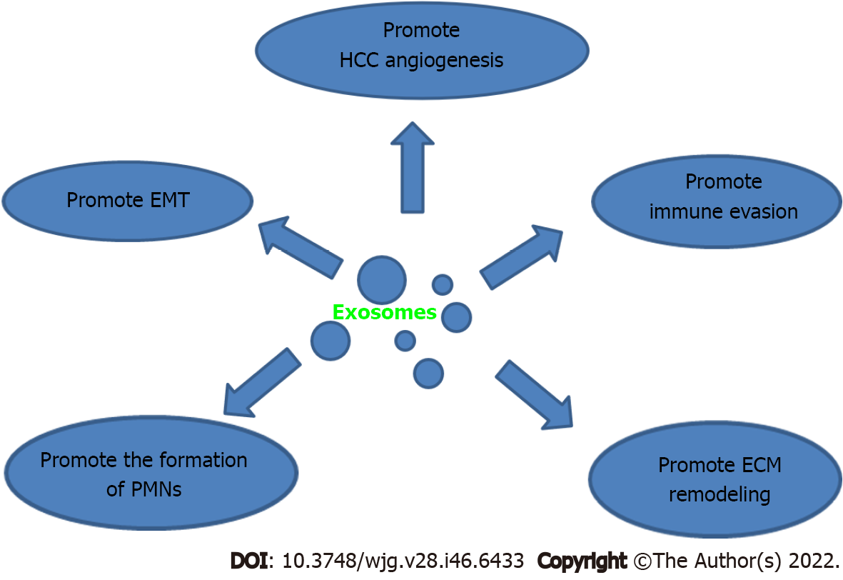
Figure 3 Schematic diagram of molecular mechanisms by which exosomes promote hepatocellular carcinoma metastasis and recurrence.
HCC: Hepatocellular carcinoma; ECM: Extracellular matrix; EMT: Epithelial mesenchymal transition; PMNs: Pre-metastatic niches.
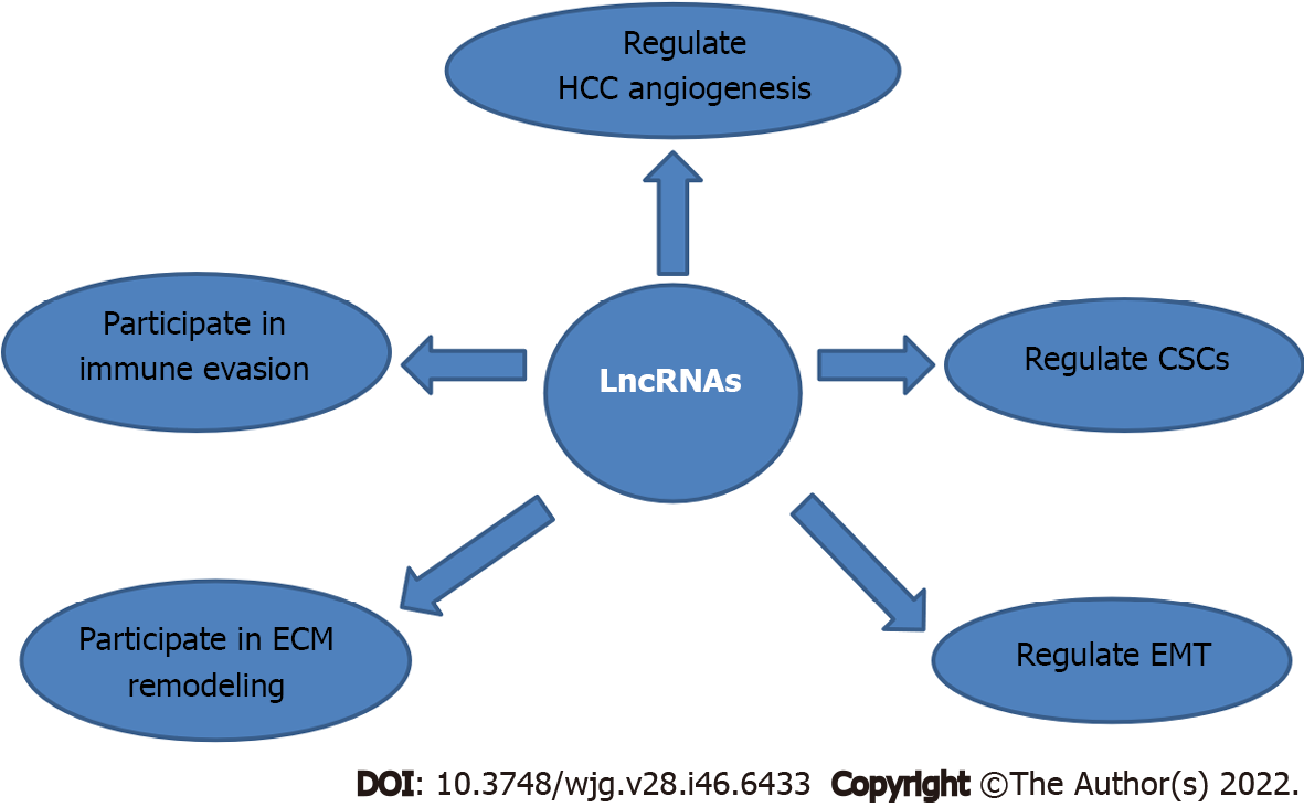
Figure 4 Schematic diagram of molecular mechanisms by which long non-coding RNAs regulate hepatocellular carcinoma metastasis and recurrence.
HCC: Hepatocellular carcinoma; LncRNAs: Long non-coding RNAs; CSCs: Cancer stem cells; ECM: Extracellular matrix; EMT: Epithelial mesenchymal transition.
- Citation: Niu ZS, Wang WH, Niu XJ. Recent progress in molecular mechanisms of postoperative recurrence and metastasis of hepatocellular carcinoma. World J Gastroenterol 2022; 28(46): 6433-6477
- URL: https://www.wjgnet.com/1007-9327/full/v28/i46/6433.htm
- DOI: https://dx.doi.org/10.3748/wjg.v28.i46.6433


