Copyright
©The Author(s) 2022.
World J Gastroenterol. Sep 28, 2022; 28(36): 5313-5323
Published online Sep 28, 2022. doi: 10.3748/wjg.v28.i36.5313
Published online Sep 28, 2022. doi: 10.3748/wjg.v28.i36.5313
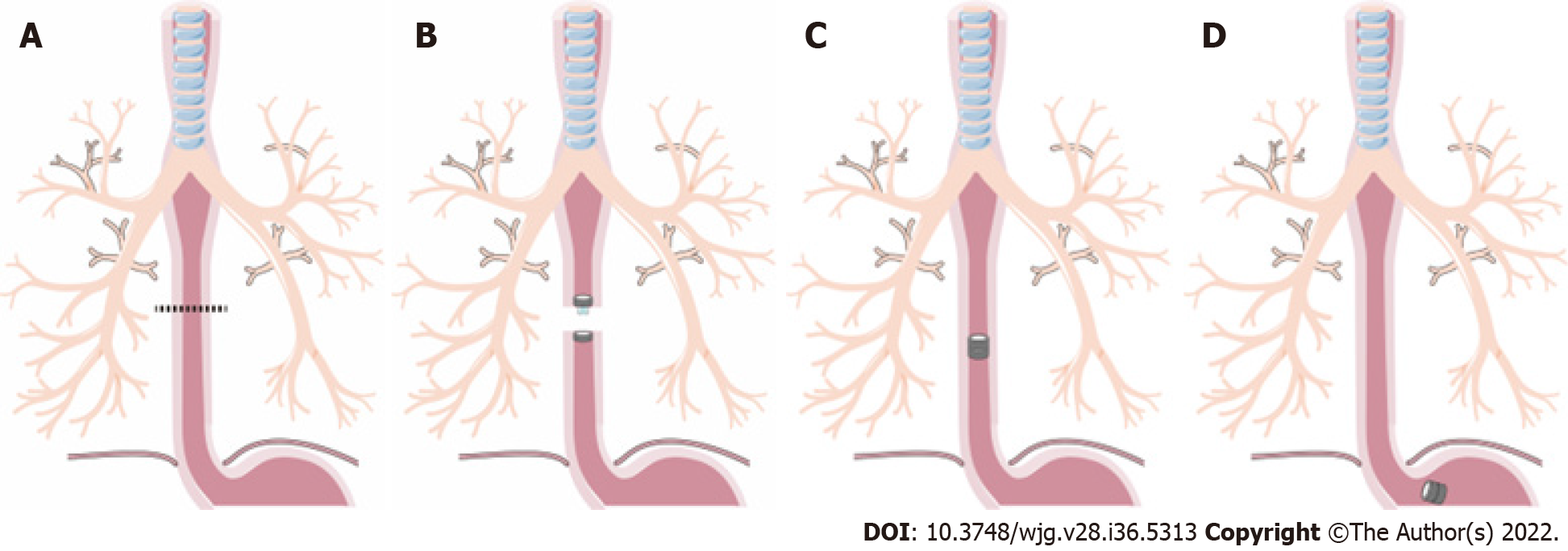
Figure 1 Surgical procedures of magnetic compress esophagus anastomosis.
A: Transecting the esophagus at the diseased region; B: Placing the parent magnetic ring (PMR) and daughter magnetic ring (DMR) on the upper and lower ends of esophagus; C: The PMR and DMR attract each other and create the anastomosis, and the esophagus reconstruction is complete, the dispositive allows food passage; D: The magnetic devices fall off the esophagus into the stomach, and the esophagus lumen is completely open.
Figure 2 The magnetic device and the appearance on X-ray, the esophageal anastomosis of magnetic compression anastomosis and hand-sewn suture.
A: The magnetic device, composed of two magnetic rings and one special catheter; B: The magnetic ring position on X-ray postoperatively, and the anastomosis face is smooth; C: Using the magnetic compression anastomosis technology to finish the esophageal anastomosis; D: The anastomosis by hand-sewn suture.
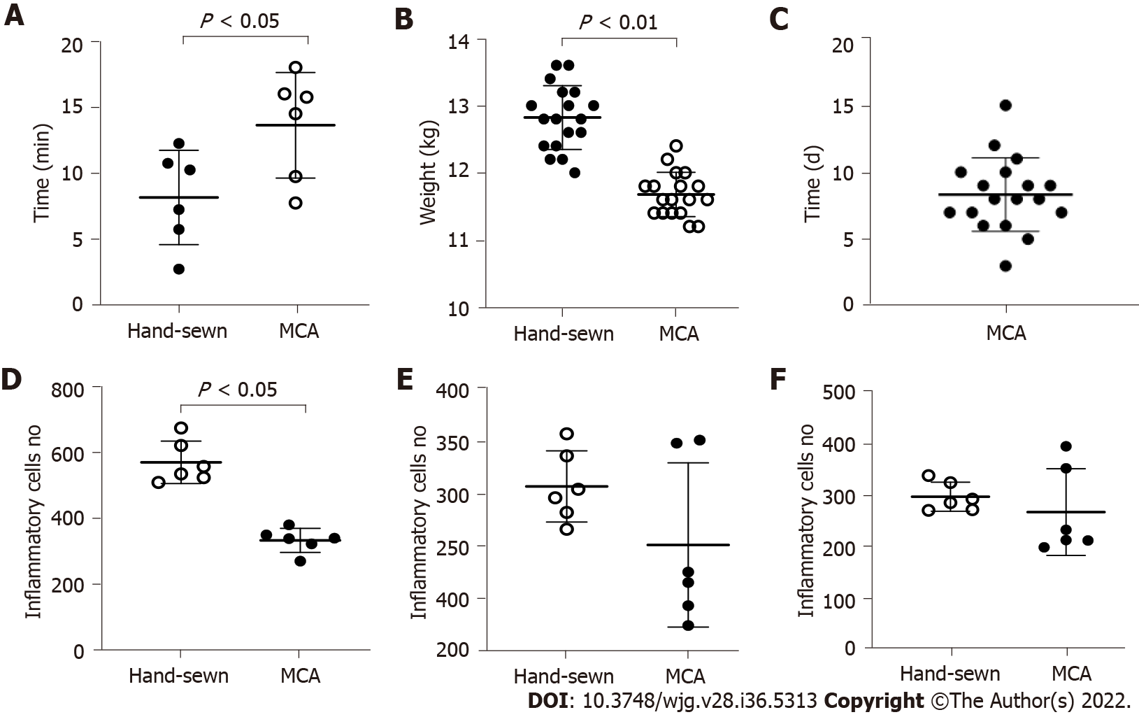
Figure 3 The analysis about anastomosis time, weight changes, the time of the magnetic device fell and the number of inflammatory cells between two groups.
A: The anastomosis time of the magnetic compression anastomosis group and hand-sewn group. There were 18 dogs in each group; B: The dogs were weighed at 1 mo, and every group contained 6 dogs. There was a significant difference between the two groups; C: The time when the magnetic device had fallen into the stomach of 18 dogs; D-F: The number of inflammatory cells respectively at 1 mo, 3 mo and 6 mo. MCA: Magnetic compression anastomosis.
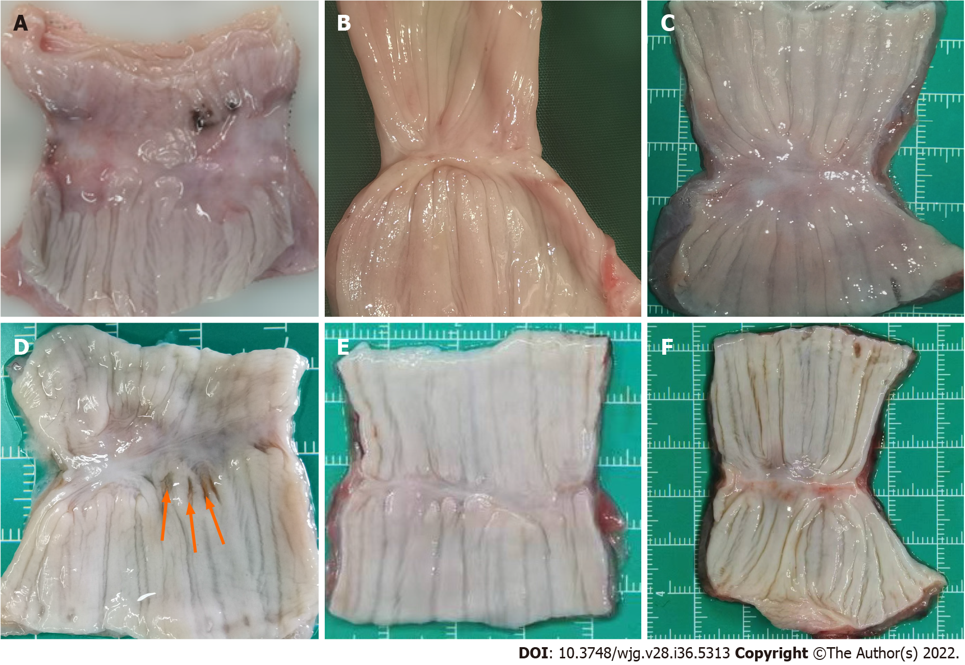
Figure 4 Gross appearance of the anastomosis in magnetic compression anastomosis and hand-sewn group.
A: The tissue of magnetic compression anastomosis (MCA) group is thin at 1 mo, but the mucous membrane is intact; B: Anastomotic tissue of the MCA group at 3 mo; C: At 6 mo, the tissue becomes smooth and flat in the MCA group. There is little difference from normal tissue; D: The tissue of the hand-sewn anastomotic stoma is incomplete, and the mucous membrane has a small pinhole at 1 mo, which was caused by the suture needle. Red arrow indicates; E: Anastomotic tissue of the hand-sewn group at 3 mo; F: Anastomotic tissue of the hand-sewn group at 6 mo.
Figure 5 Hematoxylin and eosin dye of the anastomotic tissue in magnetic compression anastomosis and hand-sewn group.
Each image consists of two parts, one with a 2 × objective view and the other with a 40 × objective view. A: The anastomotic tissue of the magnetic compression anastomosis (MCA) group at 1 mo. The mucous membrane was intact, but the muscularis was separated; B: The anastomotic tissue of the hand-sewn group at 1 mo. There were more inflammatory cells in the tissue of the hand-sewn group than that of the MCA group; C: The anastomotic tissue of the MCA group at 3 mo. The muscularis became continuous; D: The anastomotic tissue of the hand-sewn group at 3 mo; E: The anastomotic tissue of the MCA group at 6 mo; F: The anastomotic tissue of the hand-sewn group at 6 mo.
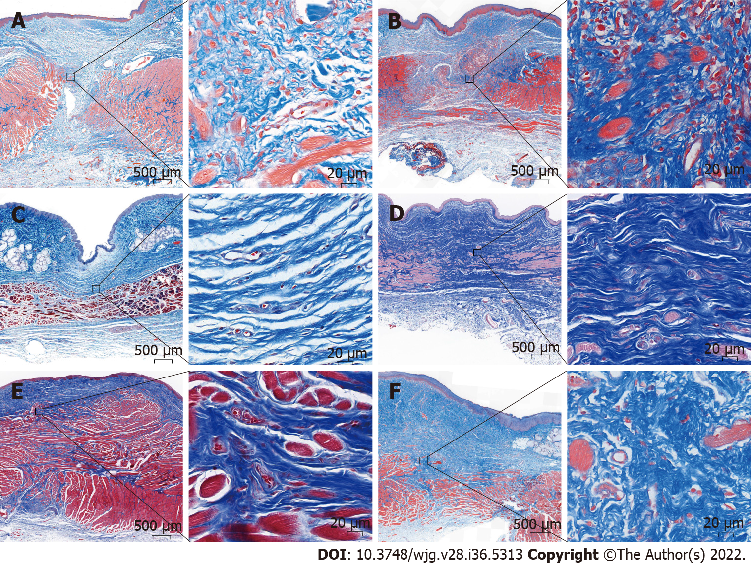
Figure 6 Masson Thrichrome dye staining of the anastomotic tissue in magnetic compression anastomosis and hand-sewn group.
Each image consists of two parts, one with a 2 × objective view and the other with a 40 × objective view. A: The anastomotic tissue of the magnetic compression anastomosis (MCA) group at 1 mo. The mucous membrane was intact, but the muscularis was separated at 1 mo, and the appearance was similar to that of hematoxylin and eosin staining; B: The anastomotic tissue of the hand-sewn group at 1 mo. There were more collagenous fibers than that in MCA group; C: The anastomotic tissue of the MCA group at 3 mo. The muscularis became continuous; D: The anastomotic tissue of the hand-sewn group at 3 mo; E: The anastomotic tissue of the MCA group at 6 mo; F: The anastomotic tissue of the hand-sewn group at 6 mo.
- Citation: Xu XH, Lv Y, Liu SQ, Cui XH, Suo RY. Esophageal magnetic compression anastomosis in dogs. World J Gastroenterol 2022; 28(36): 5313-5323
- URL: https://www.wjgnet.com/1007-9327/full/v28/i36/5313.htm
- DOI: https://dx.doi.org/10.3748/wjg.v28.i36.5313


