Copyright
©The Author(s) 2022.
World J Gastroenterol. Sep 28, 2022; 28(36): 5240-5249
Published online Sep 28, 2022. doi: 10.3748/wjg.v28.i36.5240
Published online Sep 28, 2022. doi: 10.3748/wjg.v28.i36.5240
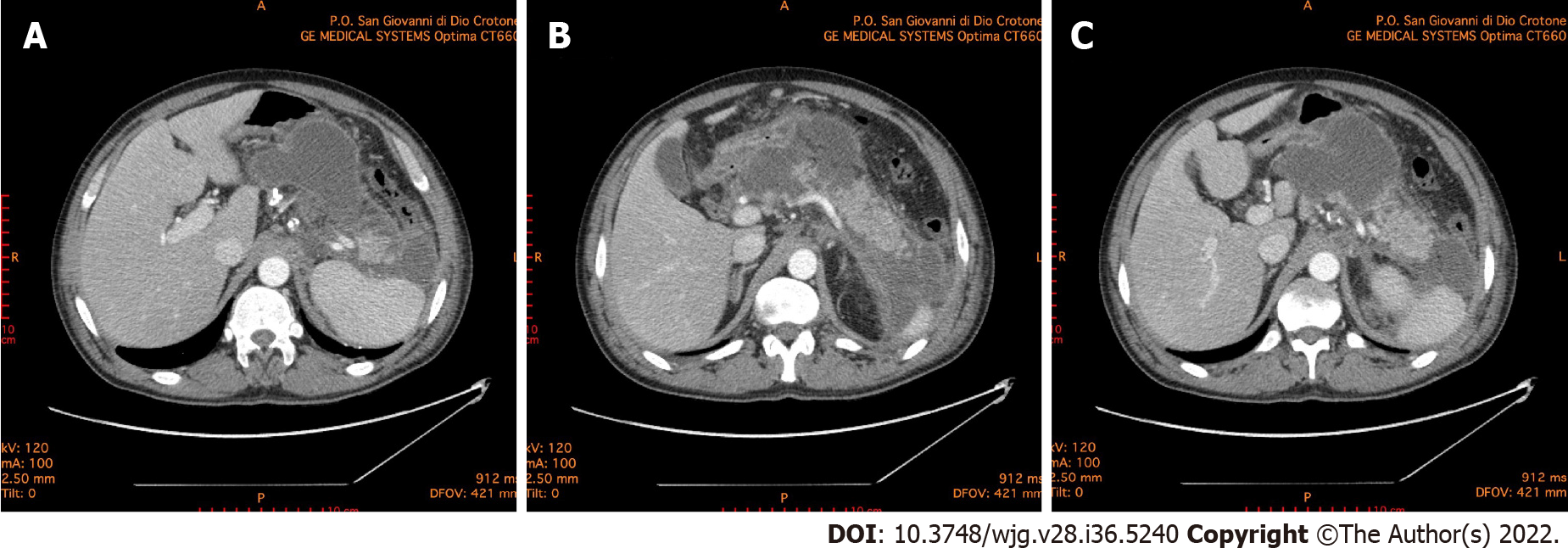
Figure 1 Abdominal computed tomography 10 d after the onset of acute pancreatitis.
A: Necrotic collection, with a noticeable wall, dislocating stomach; B: Necrotic collection occupying the majority of pancreatic head and compressing the duodenum; C: Multiple collections of fluid and necrosis involving the cephalad portion of the pancreas, multiple peripancreatic collections around the spleen and the left paracolic gutter.
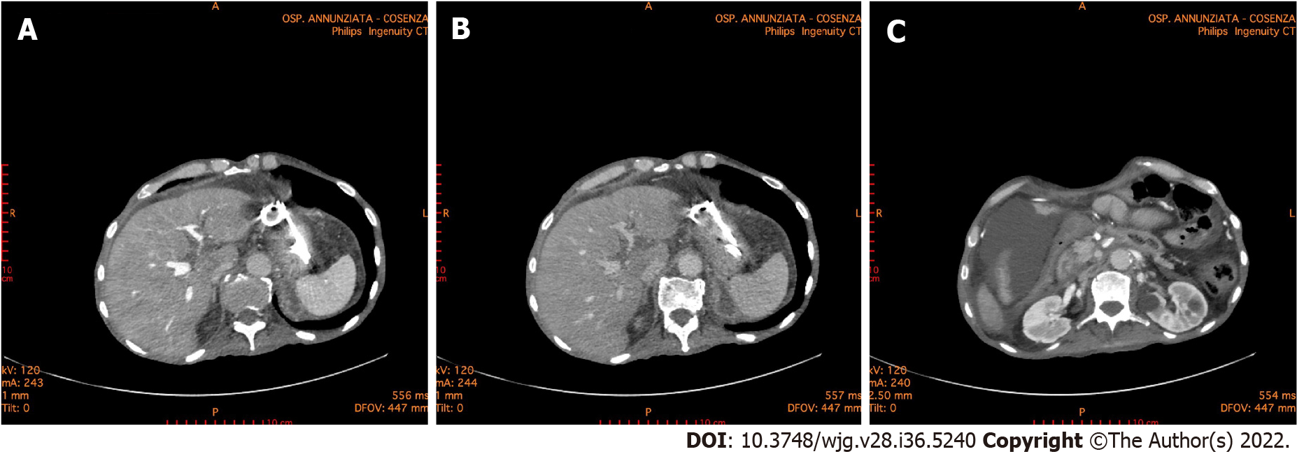
Figure 2 Abdominal computed tomography 6 mo after the onset of acute pancreatitis.
A and B: The double pigtail stents positioned during the endoscopic necrosectomy through the stomach is highlighted. Endoscopic necrosectomy through the stomach was performed 4 mo prior; C: A necrotic collection is evident in the lower right quadrant of the abdomen.
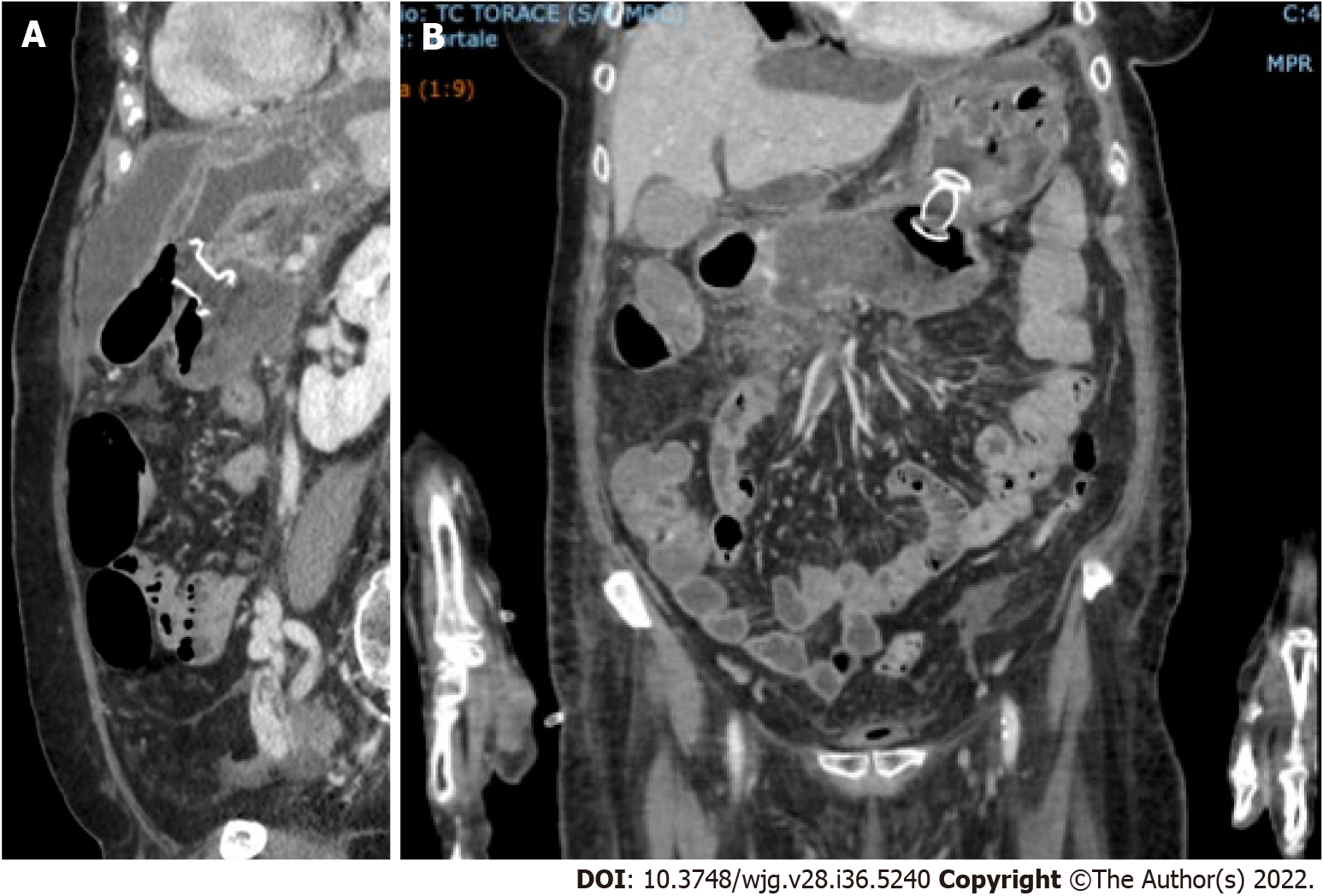
Figure 3 Abdominal computed tomography scan documenting correct placement of lumen-apposing metal stents for the treatment of walled-off necrosis in coronavirus disease 2019 positive patient.
A: Sagittal scan; B: Front scan.
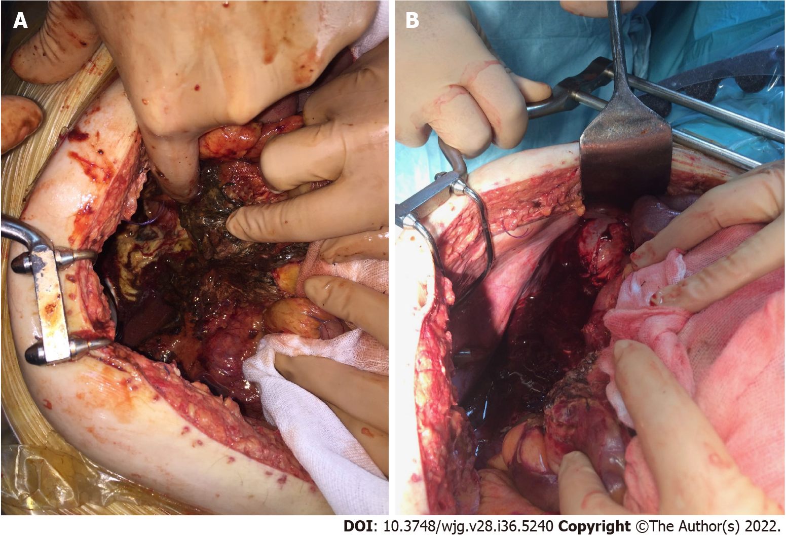
Figure 4 Intraoperative photos.
A: Surgery was performed for severe abdominal hypertension due to acute pancreatitis with necrotic collections in the retroduodenal region; B: Surgery was performed for severe abdominal hypertension due to acute pancreatitis with necrotic collections along the paracolic gutter.
- Citation: Brisinda G, Chiarello MM, Tropeano G, Altieri G, Puccioni C, Fransvea P, Bianchi V. SARS-CoV-2 and the pancreas: What do we know about acute pancreatitis in COVID-19 positive patients? World J Gastroenterol 2022; 28(36): 5240-5249
- URL: https://www.wjgnet.com/1007-9327/full/v28/i36/5240.htm
- DOI: https://dx.doi.org/10.3748/wjg.v28.i36.5240


