Copyright
©The Author(s) 2022.
World J Gastroenterol. Aug 28, 2022; 28(32): 4475-4492
Published online Aug 28, 2022. doi: 10.3748/wjg.v28.i32.4475
Published online Aug 28, 2022. doi: 10.3748/wjg.v28.i32.4475
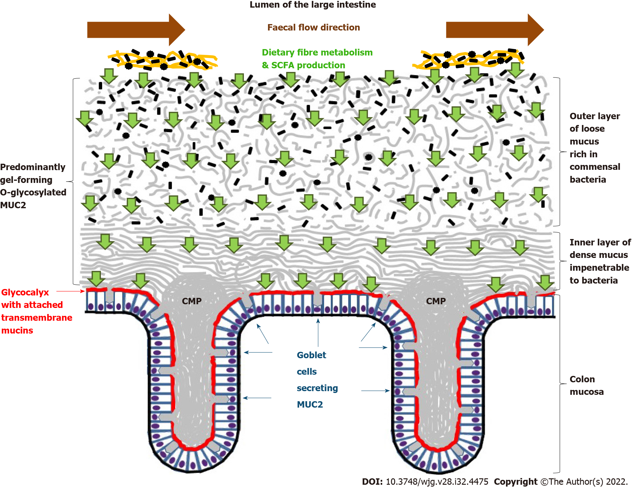
Figure 1 Schematic representation of normal human colonic mucosa and overlaying mucus layers.
Small green arrows show short-chain fatty acid transport through colon mucus layers. Small black shapes show bacteria. CMP: Colon mucus plumes; MUC2: Mucins 2; SCFA: Short-chain fatty acids.
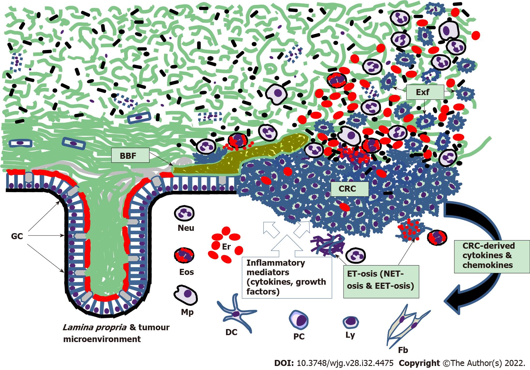
Figure 2 Scheme of colorectal mucus-associated events involved in colorectal cancer development.
BBF: Bacterial biofilm; Exf: Exfoliated malignant cells of colorectal cancer; GC: Goblet cells; Neu: Neutrophils; Eos: Eosinophils; Mp: Macrophages; Er: Erythrocytes; DC: Dendritic cells; PC: Plasma cells; Ly: Lymphocytes; Fb: Fibroblasts; ET-osis: Extracellular DNA trap formation; NET-osis: ET-osis exerted by neutrophils; EET-osis: ET-osis exerted by eosinophils. Small black shapes show bacteria; CRC: Colorectal cancer.
- Citation: Loktionov A. Colon mucus in colorectal neoplasia and beyond. World J Gastroenterol 2022; 28(32): 4475-4492
- URL: https://www.wjgnet.com/1007-9327/full/v28/i32/4475.htm
- DOI: https://dx.doi.org/10.3748/wjg.v28.i32.4475


