Copyright
©The Author(s) 2022.
World J Gastroenterol. Aug 21, 2022; 28(31): 4456-4462
Published online Aug 21, 2022. doi: 10.3748/wjg.v28.i31.4456
Published online Aug 21, 2022. doi: 10.3748/wjg.v28.i31.4456
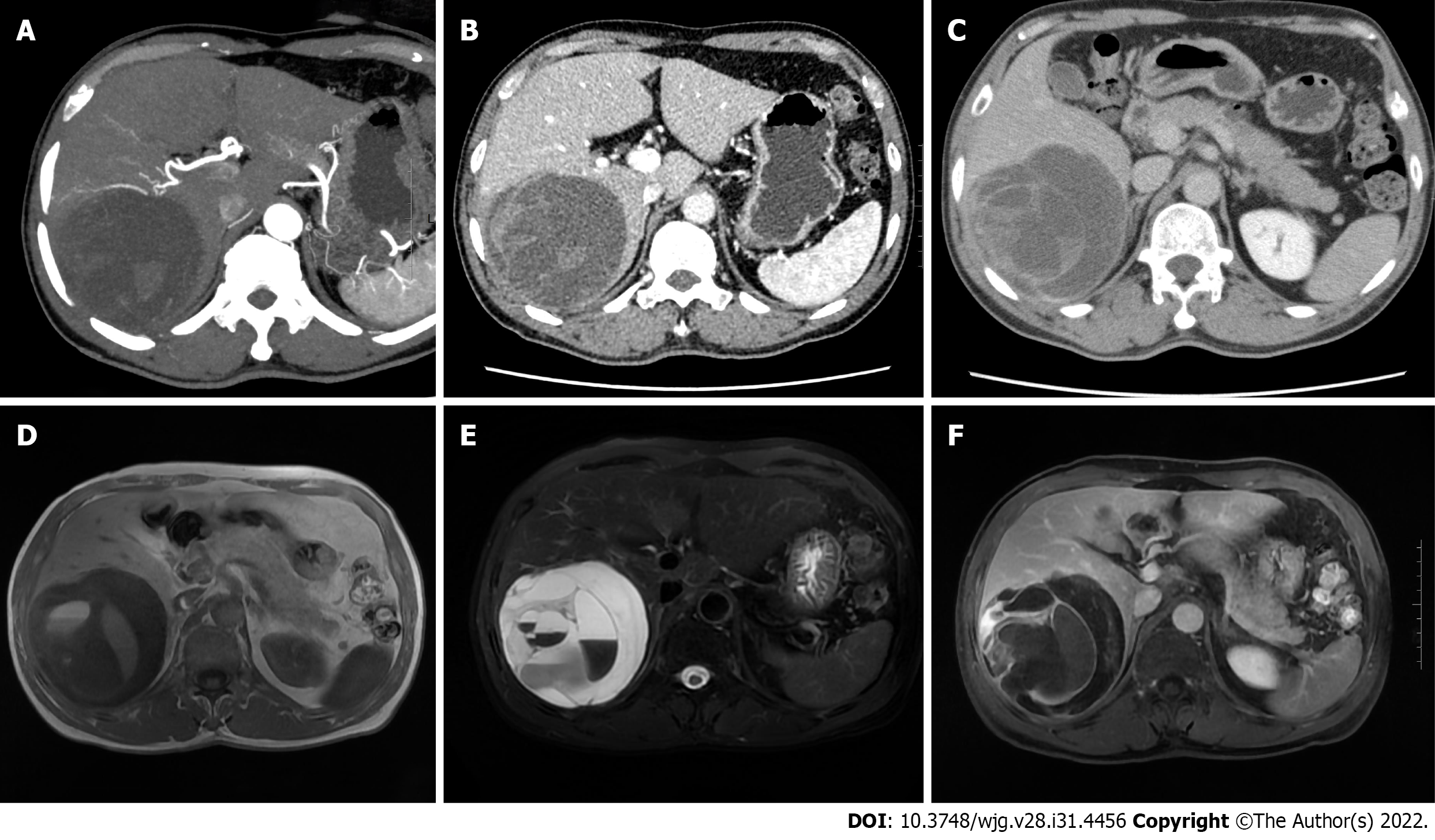
Figure 1 Abdominal contrast-enhanced computed tomography and hepatic vascular imaging examination, and magnetic resonance imaging examination.
A: The right hepatic cystic-solid mass was supplied by the right hepatic artery, and the twisting of the supplying artery was seen; B and C: The portal phase and delayed phase, respectively; the solid portion and the septum were enhanced, and the intracapsular septum was clearly displayed; D and E: The lesions were low signal on T1-weighted imaging and high signal on T2-weighted imaging, with multiple septa of uneven thickness and fluid-fluid levels; F: After enhancement, the solid components and septa of the mass were significantly enhanced, but the cystic components were not.
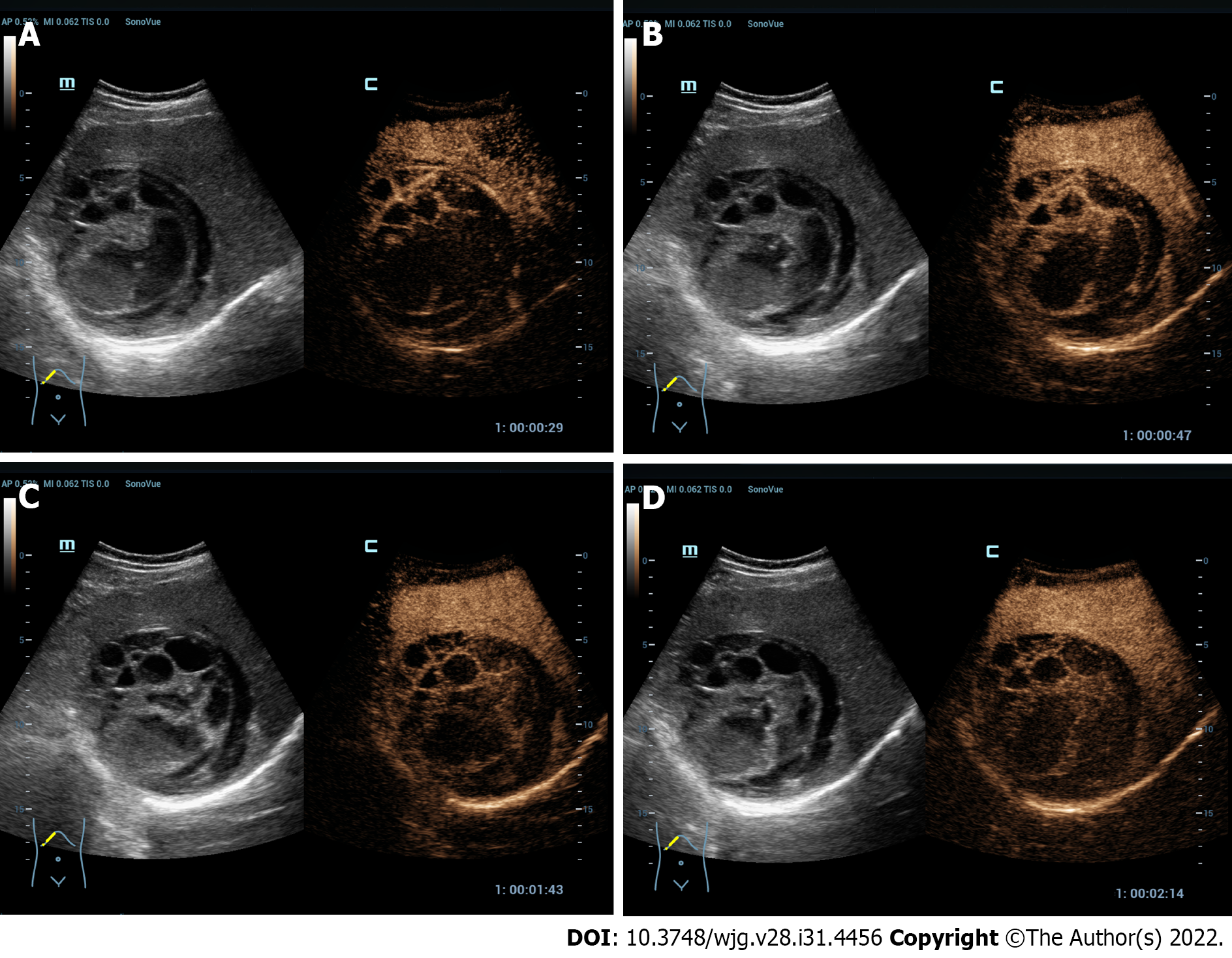
Figure 2 Contrast-enhanced ultrasound examination.
The peripheral parenchyma and internal septa of the mixed-echo mass showed hyper-enhancement in the arterial phase, and iso-enhancement in the portal and delayed phases. A: Arterial phase; B: Early portal phase; C: Late portal phase; D: Delayed phase.
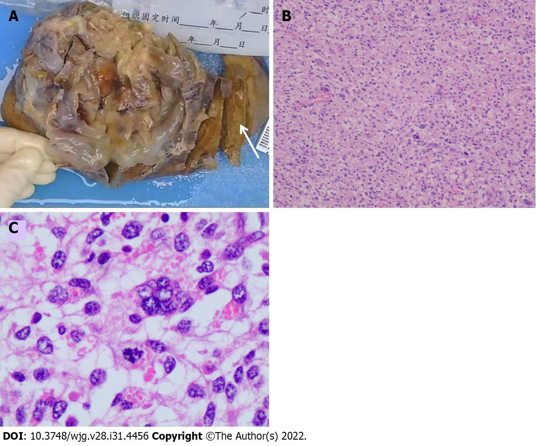
Figure 3 Gross pathology specimen and pathological microscopy.
A: A cystic-solid mass was seen with the naked eye, with soft texture and inconspicuous tumor capsule; its cut surface was polycystic, with yellowish jelly-like substances inside the cysts, and the cyst wall thickness was 1 mm-12 mm; no cirrhotic changes were observed in the peripheral liver tissue (arrow); B and C: Spindle-shaped cells are arranged in fascicles under light microscope, eosinophilic cytoplasm with atypia and mitotic figures were seen.
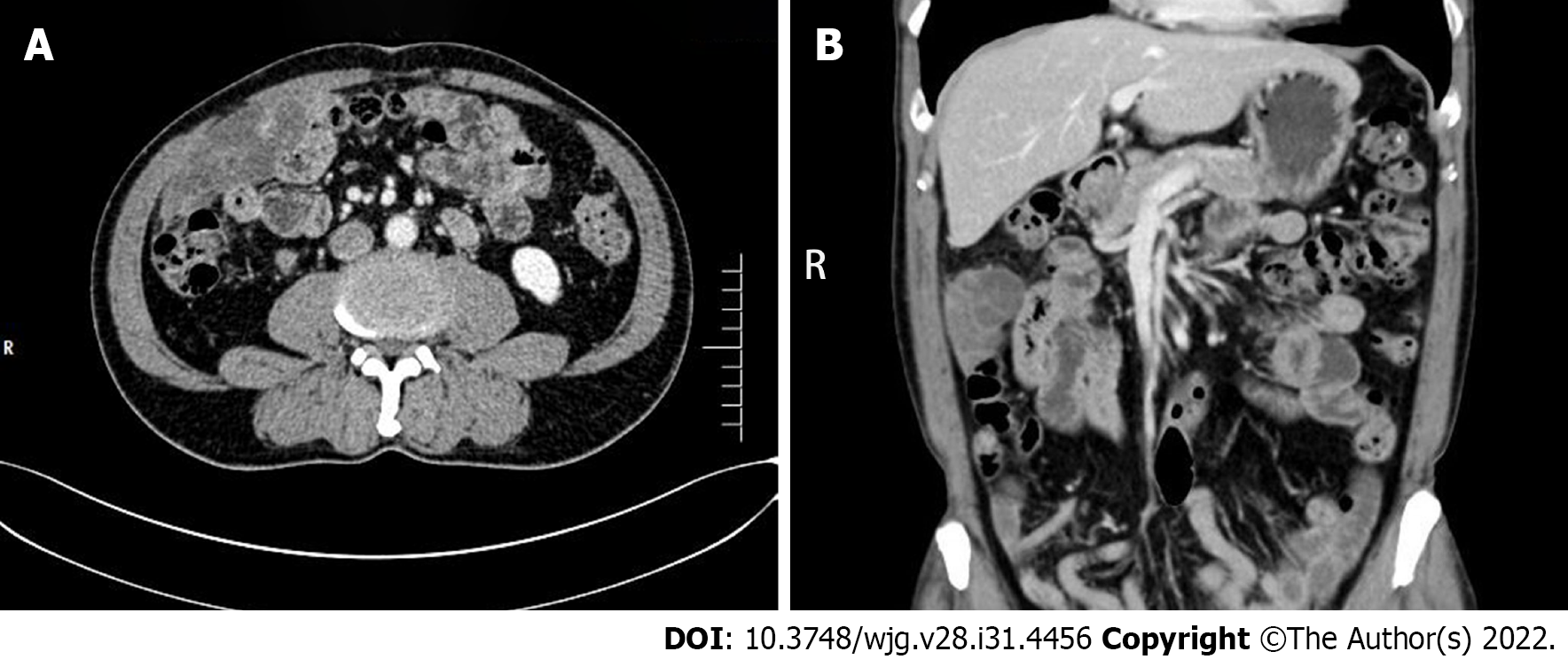
Figure 4 Abdominal contrast-enhanced computed tomography examination at 7-mo follow-up.
A and B: The right middle abdomen of the patient in contrast-enhanced computed tomography showed a cystic-solid lesion, with the similar property as a liver lesion. After enhancement, the mass wall was enhanced.
- Citation: Li J, Huang XY, Zhang B. Low-grade myofibroblastic sarcoma of the liver misdiagnosed as cystadenoma: A case report. World J Gastroenterol 2022; 28(31): 4456-4462
- URL: https://www.wjgnet.com/1007-9327/full/v28/i31/4456.htm
- DOI: https://dx.doi.org/10.3748/wjg.v28.i31.4456


