Copyright
©The Author(s) 2022.
World J Gastroenterol. Jul 28, 2022; 28(28): 3620-3626
Published online Jul 28, 2022. doi: 10.3748/wjg.v28.i28.3620
Published online Jul 28, 2022. doi: 10.3748/wjg.v28.i28.3620
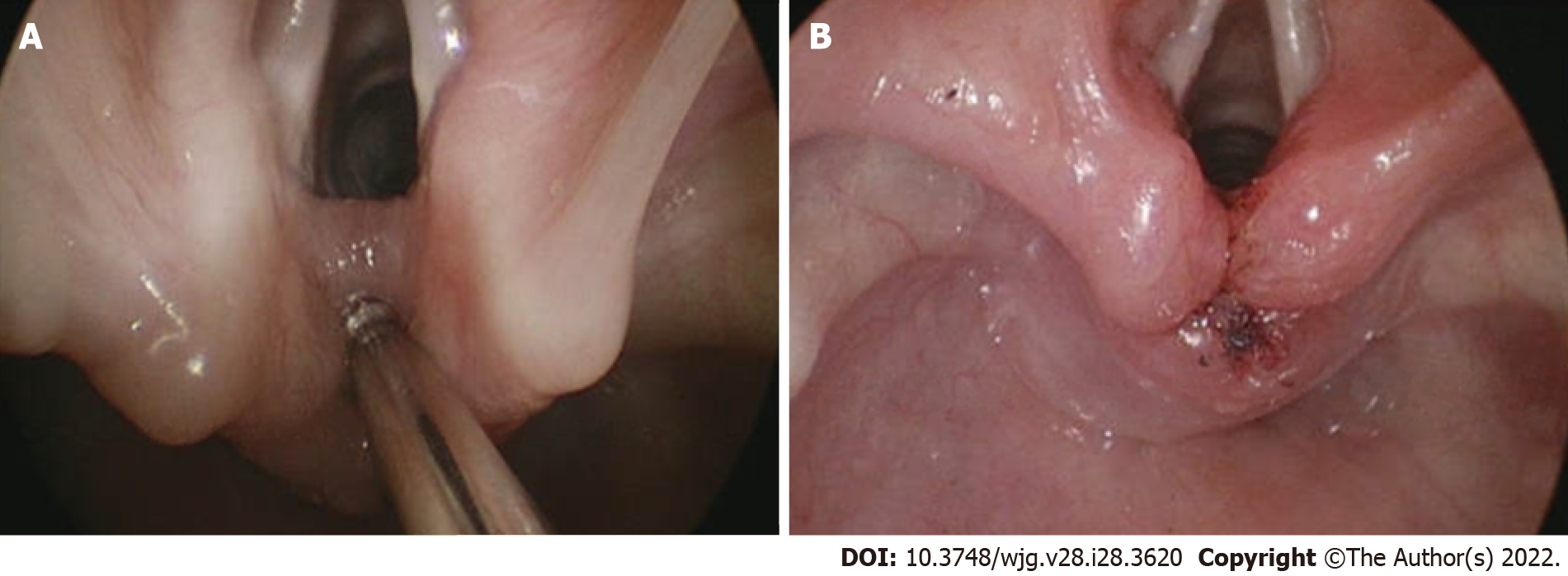
Figure 1 Endoscopic assessment and management for a laryngeal cleft which can contribute to aspiration.
A: Probing of interaytenoid region to assess for cleft; B: Post laryngeal cleft repair using laser and suturing.
Figure 2 Endoscopic grading of subglottic stenosis.
A: Grade I: 0%-50% obstruction; B: Grade II: 51%-70% obstruction; C: Grade III: 71%-99%; D: Grade IV: No detectable lumen.
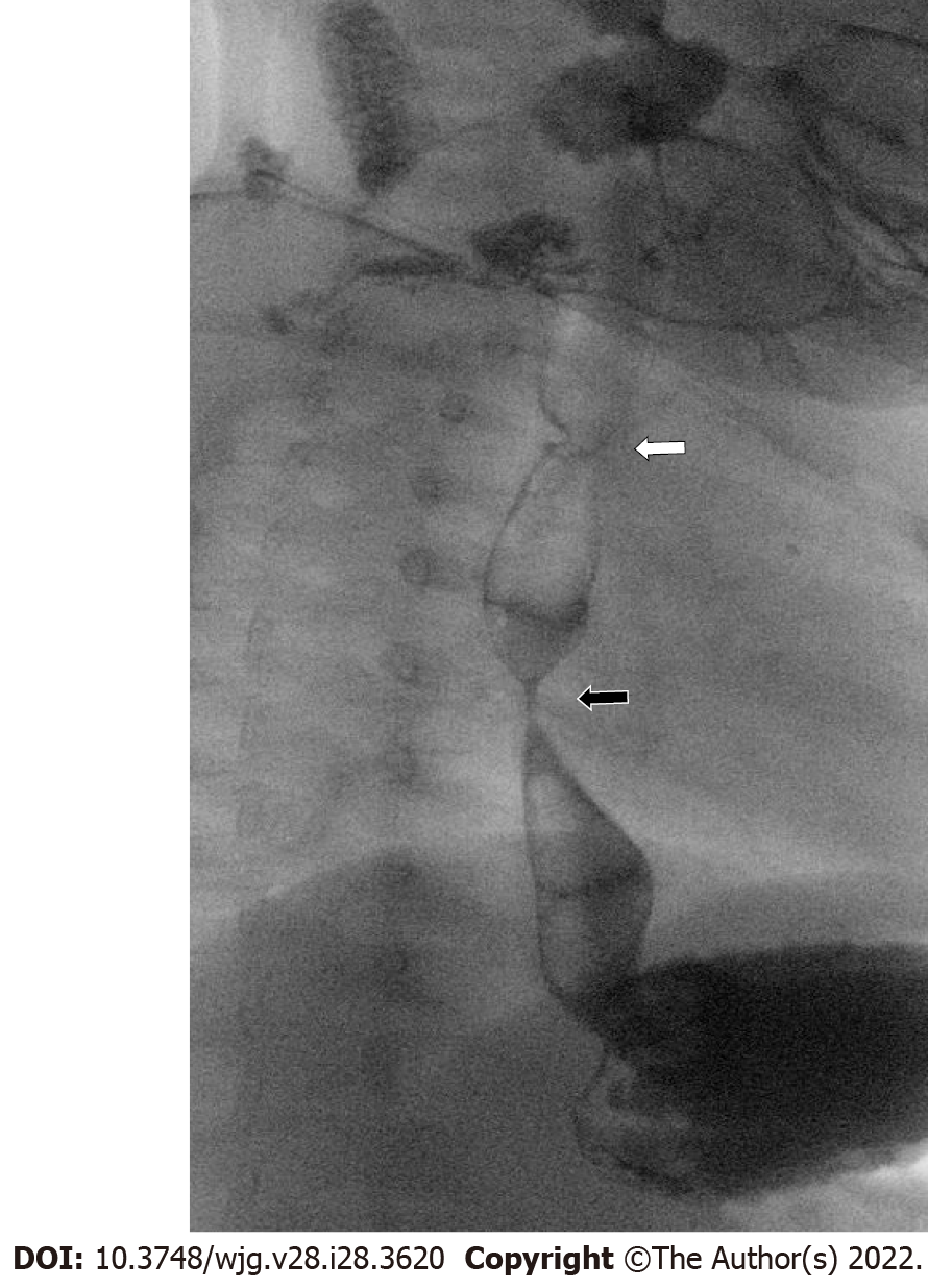
Figure 3 Fluoroscopic esophageal evaluation in high-risk patients.
Esophagram in 10-month-old with repaired esophageal atresia presenting with feeding difficulty and poor growth, showing previously unrecognized distal esophageal congenital stricture (black arrow), far below the surgical repair site (white arrow).
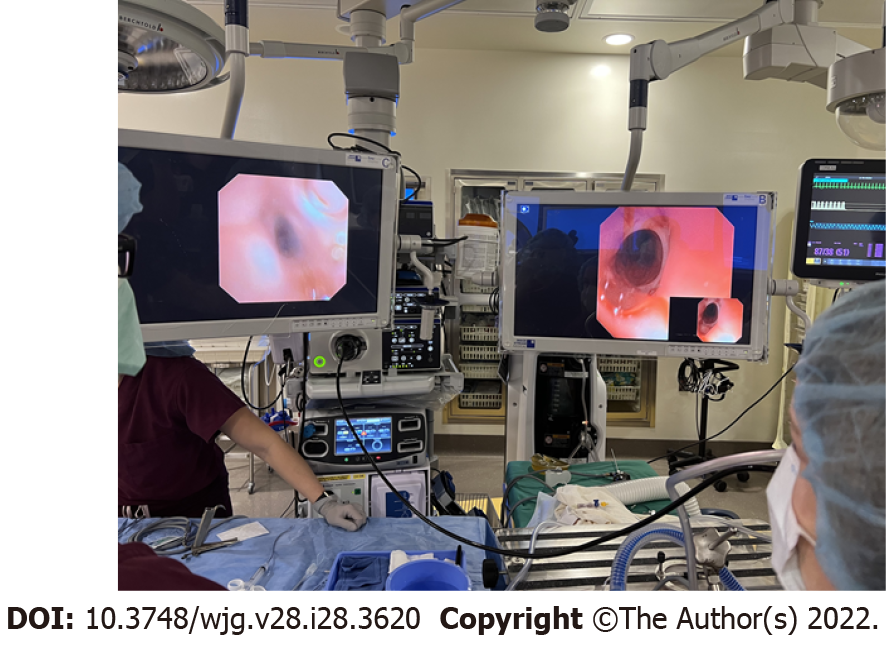
Figure 4 Simultaneous tracheal and esophageal endoscopy in a patient with history of esophageal atresia to assess for persistent tracheoesophageal fistula.
- Citation: Kanotra SP, Weiner R, Rahhal R. Making the case for multidisciplinary pediatric aerodigestive programs. World J Gastroenterol 2022; 28(28): 3620-3626
- URL: https://www.wjgnet.com/1007-9327/full/v28/i28/3620.htm
- DOI: https://dx.doi.org/10.3748/wjg.v28.i28.3620


