Copyright
©The Author(s) 2022.
World J Gastroenterol. Jul 28, 2022; 28(28): 3555-3572
Published online Jul 28, 2022. doi: 10.3748/wjg.v28.i28.3555
Published online Jul 28, 2022. doi: 10.3748/wjg.v28.i28.3555
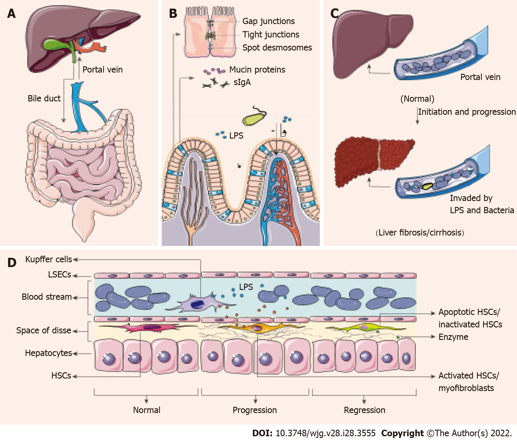
Figure 1 Mechanism of gut microbiota-related liver fibrosis/cirrhosis.
A: Gut-liver axis. The close bidirectional connection between gut and liver is mainly through the portal vein and bile duct; B: Intestinal barriers. From the intestine lumen, intestinal barriers are mainly formed by mucin proteins, sIgA and intercellular junctions, especially tight junctions between intestinal epithelial cells. The asterisk means when the intestinal barriers are weakened or broken, microbe/antigen translocation ensues; C: Liver fibrosis/cirrhosis and gut microbe/antigen translocation. Compared with normal state, gut microbe/antigen translocation and liver fibrosis/cirrhosis may drive each other in chronic hepatitis B patients; D: Mechanisms of liver fibrosis/cirrhosis process and regression. Receiving the activation signal, hepatic stellate cells (HSCs) are activated into fibroblasts to form the fiber. As the activation signal ceases, the activated HSCs are inactivated or apoptotic. When fiber degradation predominated, fibrosis is reversed. HSCs: Hepatic stellate cells; LPS: Lipopolysaccharide; LSECs: Liver sinusoidal endothelial cells.
- Citation: Li YG, Yu ZJ, Li A, Ren ZG. Gut microbiota alteration and modulation in hepatitis B virus-related fibrosis and complications: Molecular mechanisms and therapeutic inventions. World J Gastroenterol 2022; 28(28): 3555-3572
- URL: https://www.wjgnet.com/1007-9327/full/v28/i28/3555.htm
- DOI: https://dx.doi.org/10.3748/wjg.v28.i28.3555


