Copyright
©The Author(s) 2022.
World J Gastroenterol. Jul 14, 2022; 28(26): 3258-3273
Published online Jul 14, 2022. doi: 10.3748/wjg.v28.i26.3258
Published online Jul 14, 2022. doi: 10.3748/wjg.v28.i26.3258
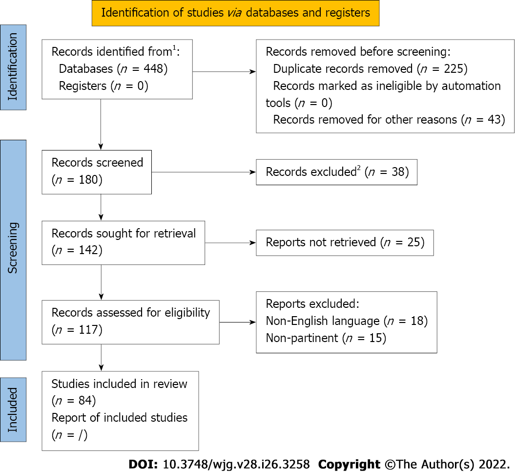
Figure 1 The flow chart showing the process of study selection.
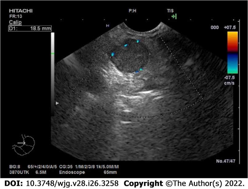
Figure 2 Endoscopic appearance at endoscopic ultrasound of a pancreatic neuroendocrine neoplasm with marginal vascularization.
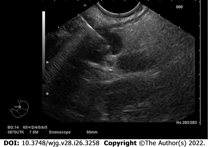
Figure 3 Endoscopic appearance at endoscopic ultrasound of a pancreatic neuroendocrine neoplasm located at the tail of the pancreas during fine needle aspiration/biopsy procedure.
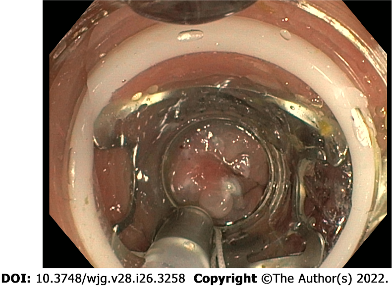
Figure 4 Over-the-scope clipping system for endoscopic full-thickness resection of a rectal neuroendocrine neoplasm.
- Citation: Rossi RE, Elvevi A, Gallo C, Palermo A, Invernizzi P, Massironi S. Endoscopic techniques for diagnosis and treatment of gastro-entero-pancreatic neuroendocrine neoplasms: Where we are. World J Gastroenterol 2022; 28(26): 3258-3273
- URL: https://www.wjgnet.com/1007-9327/full/v28/i26/3258.htm
- DOI: https://dx.doi.org/10.3748/wjg.v28.i26.3258


