Copyright
©The Author(s) 2022.
World J Gastroenterol. Jul 14, 2022; 28(26): 3101-3115
Published online Jul 14, 2022. doi: 10.3748/wjg.v28.i26.3101
Published online Jul 14, 2022. doi: 10.3748/wjg.v28.i26.3101
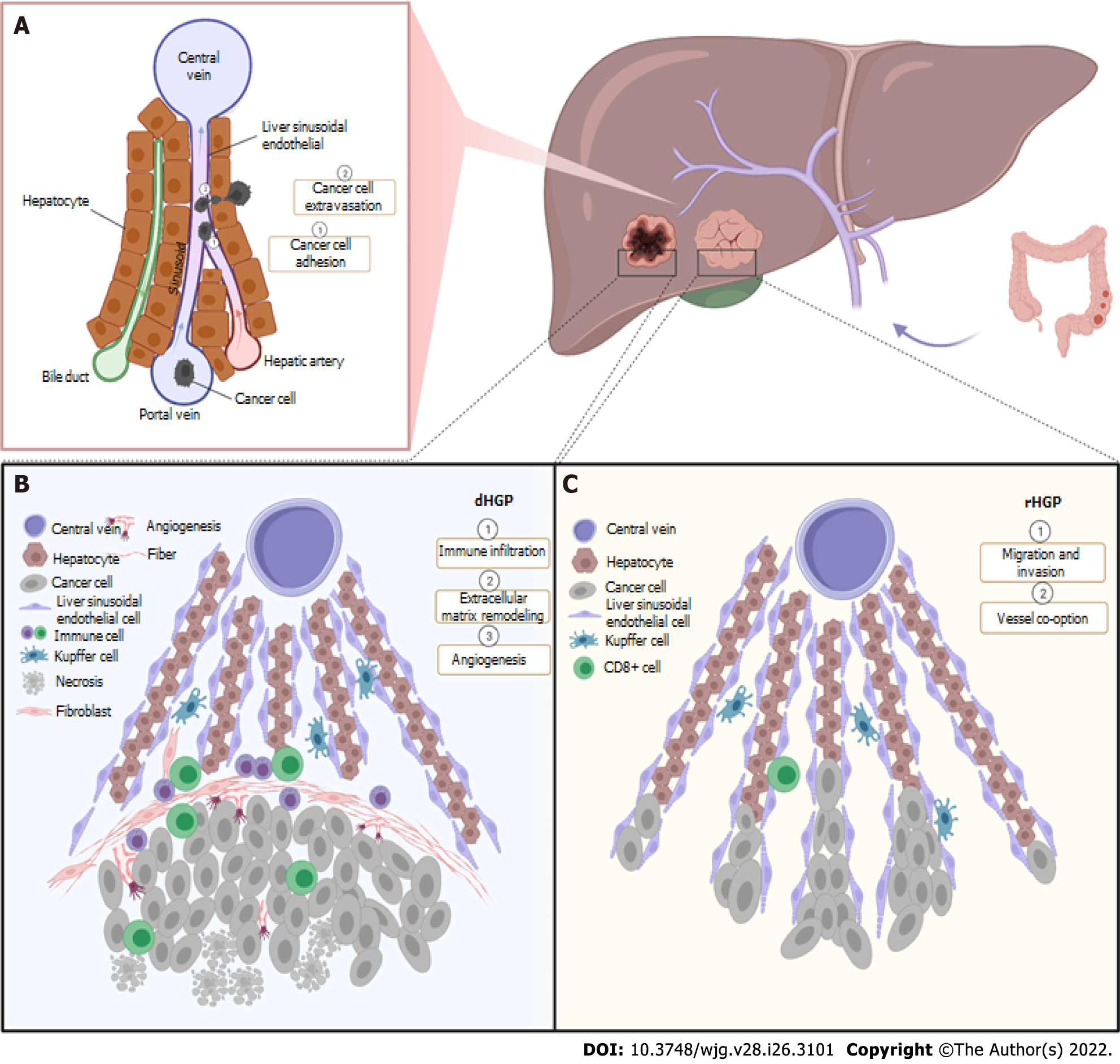
Figure 1 Formation and mechanism of desmoplastic histopathological growth pattern and replacement histopathological growth pattern.
A: Cancer cells originating from colorectal cancer arrive in the liver via portal vein, adhere to lumen of liver sinusoid and migrate with extravasation through fenestrae on liver sinusoidal endothelial cells (LSECs); B: There is a desmoplastic rim in interface of tumor with desmoplastic histopathological growth pattern, with tumor cells destroying liver plate, causing immune infiltration and extracellular matrix remodeling induced by activated fibroblasts and deposited fiber. Both angiogenesis and necrosis are presented in the tumor; C: In replacement histopathological growth pattern, tumor cells with highly migration and invasion replace hepatocytes and co-opt LSECs but without disturbing the liver structure and extensive immune infiltration. dHGP: Desmoplastic histopathological growth pattern; rHGP: Replacement histopathological growth pattern.
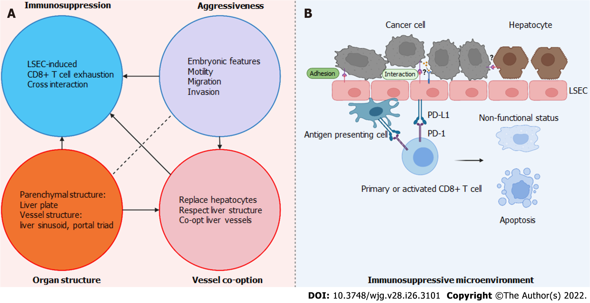
Figure 2 “Advance under camouflage” hypothesis for replacement histopathological growth pattern.
A: “Advance under camouflage” hypothesis of replacement histopathological growth pattern (rHGP) includes four elements. Embryonic features, motility, migration and adhesion contributes to aggressiveness of tumor, which drives tumor progression as an intrinsic factor. Tumor cells interact with liver sinusoidal endothelial cells (LSECs) and hepatocytes, so that tumor cells educate these normal liver cells and remodel the immune microenvironment into a tolerance state via CD8+ T cells. In this manner, under camouflage of LSECs, tumor cells are able to survive and slowly progress. Co-opting LSECs enable the tumor to obtain sufficient blood supply and less chance of exposure to immune system. With its unique parenchymal and vessel structure, organ architecture of liver supports the whole advance process; B: Immunosuppressive microenvironment in tumor with rHGP. Under cross interaction of cancer cells and LSECs and other immune cells (unknown mechanism), programmed death (PD) ligand 1 on LSECs and antigen presenting cells binds to PD-1 on T cells making CD8+ T cells activated to a dynamic but nonfunctional type which fails to produce effector cytokines and has decreased cellular cytotoxicity, or is apoptostic. LSEC: Liver sinusoidal endothelial cell; PD: Programmed death.
- Citation: Kong BT, Fan QS, Wang XM, Zhang Q, Zhang GL. Clinical implications and mechanism of histopathological growth pattern in colorectal cancer liver metastases. World J Gastroenterol 2022; 28(26): 3101-3115
- URL: https://www.wjgnet.com/1007-9327/full/v28/i26/3101.htm
- DOI: https://dx.doi.org/10.3748/wjg.v28.i26.3101


