Copyright
©The Author(s) 2022.
World J Gastroenterol. Jun 14, 2022; 28(22): 2482-2493
Published online Jun 14, 2022. doi: 10.3748/wjg.v28.i22.2482
Published online Jun 14, 2022. doi: 10.3748/wjg.v28.i22.2482
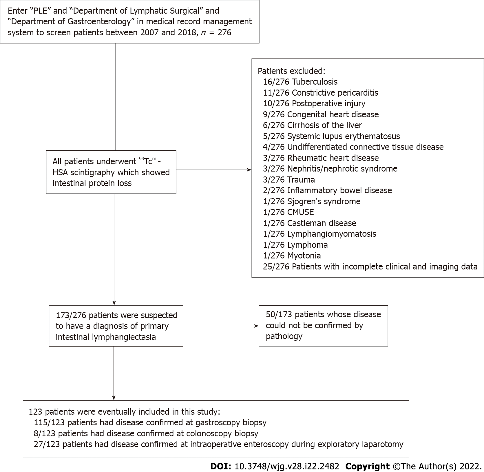
Figure 1 Patient inclusion flowchart showing the number of patients, radionuclide imaging, digestive endoscopy evaluation, and pathologic analysis.
CMUSE: Cryptogenic multifocal ulcerous stenosing enteritis; PLE: Protein-losing enteropathy.
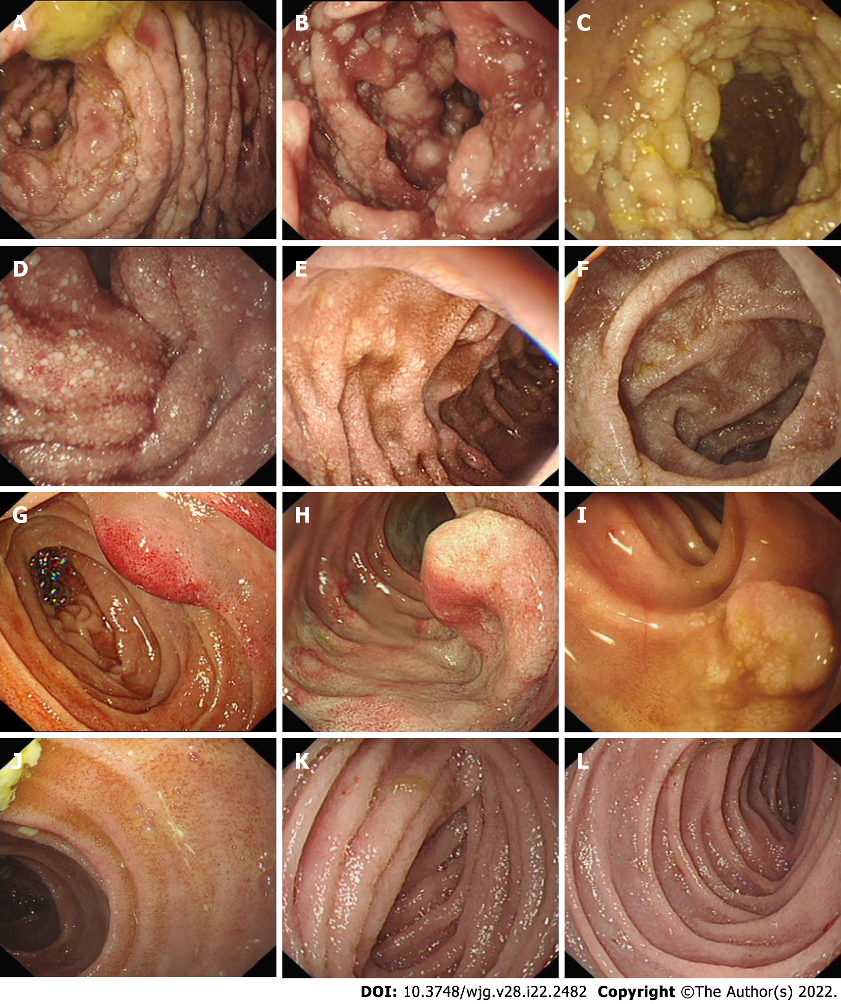
Figure 2 Endoscopic images of patients with primary intestinal lymphangiectasia.
The images show white mucosal plaques and spots in the small intestine, divided into four types. A-C: Nodular type; D-F: Granular type; G-I: Vesicular type; J-L: Edematous type.
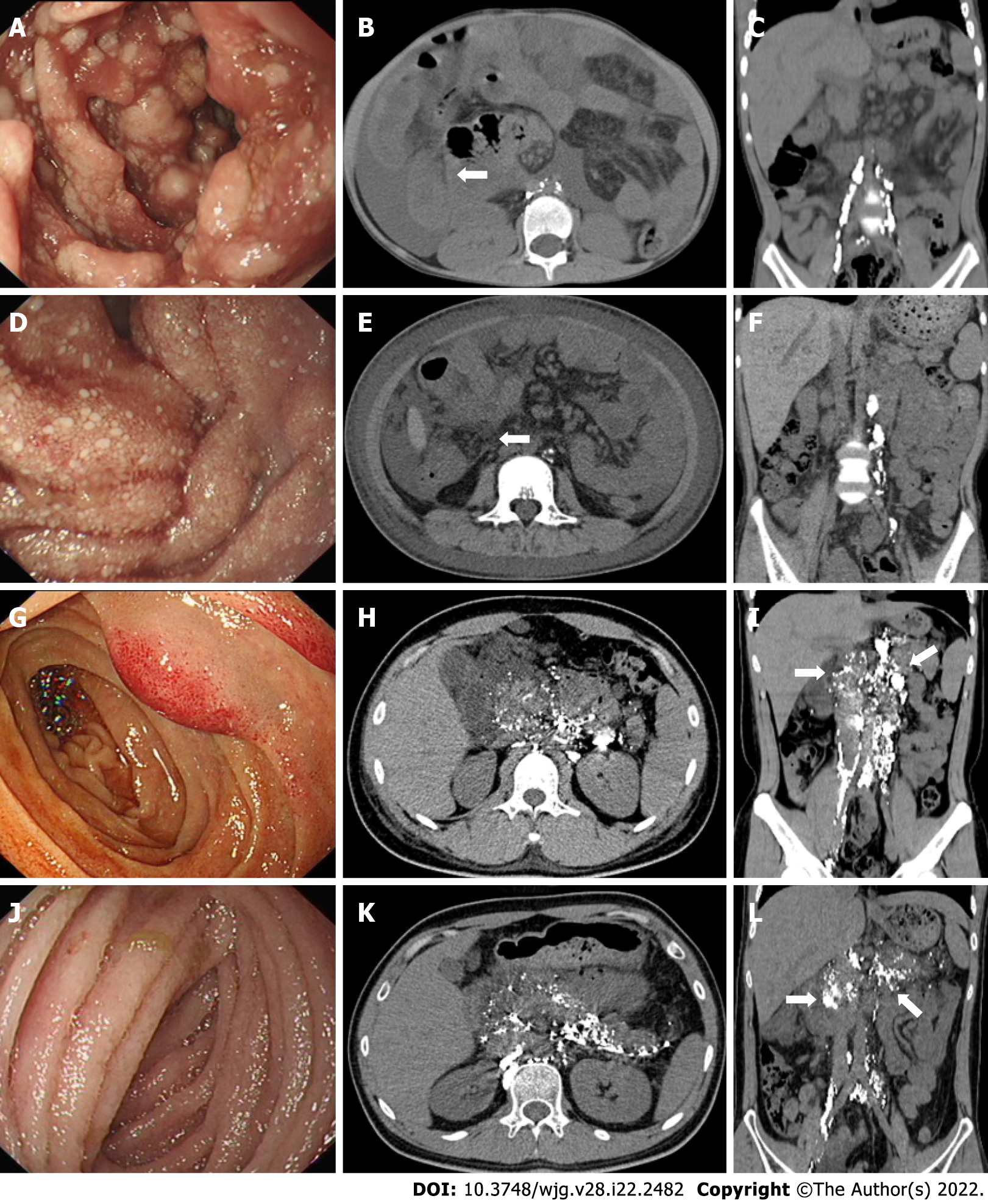
Figure 3 Post-lymphographic computed tomography images corresponding to the four types of primary intestinal lymphangiectasia under endoscopy.
A-C: Nodular type: The post-lymphographic computed tomography (PLCT) corresponding to nodular type indicates the intestinal wall thickening of the small intestine (white arrows), increased mesenteric density, and no contrast agent distribution in the mesentery and intestinal wall; D-F: Granular type: The PLCT corresponding to granular type indicates that the intestinal wall annular thickening of the small intestine, increased mesenteric density (white arrows), and no contrast agent distribution in the mesentery and intestinal wall; G-I: Vesicular type: The PLCT corresponding to vesicular type indicates the distribution of contrast agents in the intestinal wall, mesentery, peripancreas, hilar area of the liver, and retroperitoneum (white arrows), suggesting lymphatic dilation and abnormal distribution in the above areas; J-L: Edematous type: The PLCT corresponding to edematous type indicates the distribution of contrast agents in the intestinal wall, mesentery, peripancreas, gallbladder fossa, hilar area of the liver, and retroperitoneum (white arrows).
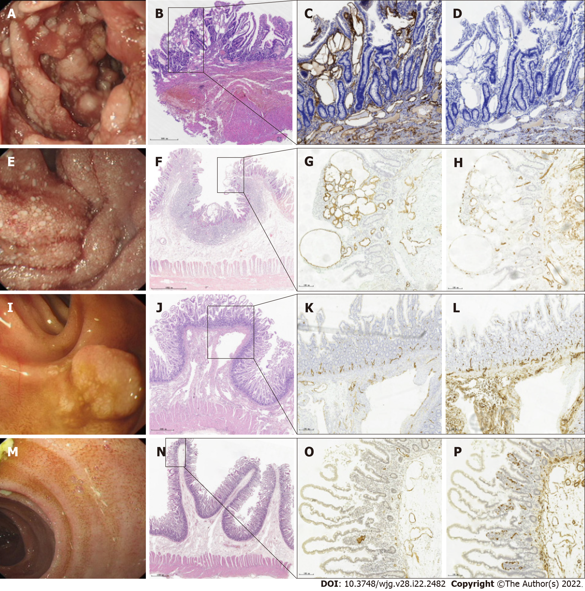
Figure 4 Endoscopic images and corresponding histological findings of primary intestinal lymphangiectasia.
A, E, I, and M: Four types of endoscopic images in patients with primary intestinal lymphangiectasia; B, F, J, and N: Hematoxylin and eosin stain showing the full mucosa of the small intestine; C, G, K, and O: Immunohistochemical stain shows that D2-40 positive cells are located in the cytoplasm and envelope of lymphatic epithelial cells and are brownish yellow (magnification: × 10), indicating dilated lymphatics; D, H, L, and P: CD34 stain showing normal vascular endothelial cells (magnification: × 10).
Figure 5 Four patterns of mucosal findings under endoscopy.
Type I: Nodular type; Type II: Granular type; Type III: Vesicular type; Type IV: Edematous type.
- Citation: Meng MM, Liu KL, Xue XY, Hao K, Dong J, Yu CK, Liu H, Wang CH, Su H, Lin W, Jiang GJ, Wei N, Wang RG, Shen WB, Wu J. Endoscopic classification and pathological features of primary intestinal lymphangiectasia. World J Gastroenterol 2022; 28(22): 2482-2493
- URL: https://www.wjgnet.com/1007-9327/full/v28/i22/2482.htm
- DOI: https://dx.doi.org/10.3748/wjg.v28.i22.2482


