Copyright
©The Author(s) 2022.
World J Gastroenterol. Jun 7, 2022; 28(21): 2350-2360
Published online Jun 7, 2022. doi: 10.3748/wjg.v28.i21.2350
Published online Jun 7, 2022. doi: 10.3748/wjg.v28.i21.2350
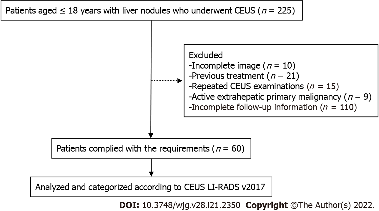
Figure 1 Flow diagram for the study population.
CEUS: Contrast-enhanced ultrasound; LI-RADS: Liver imaging reporting and data system.
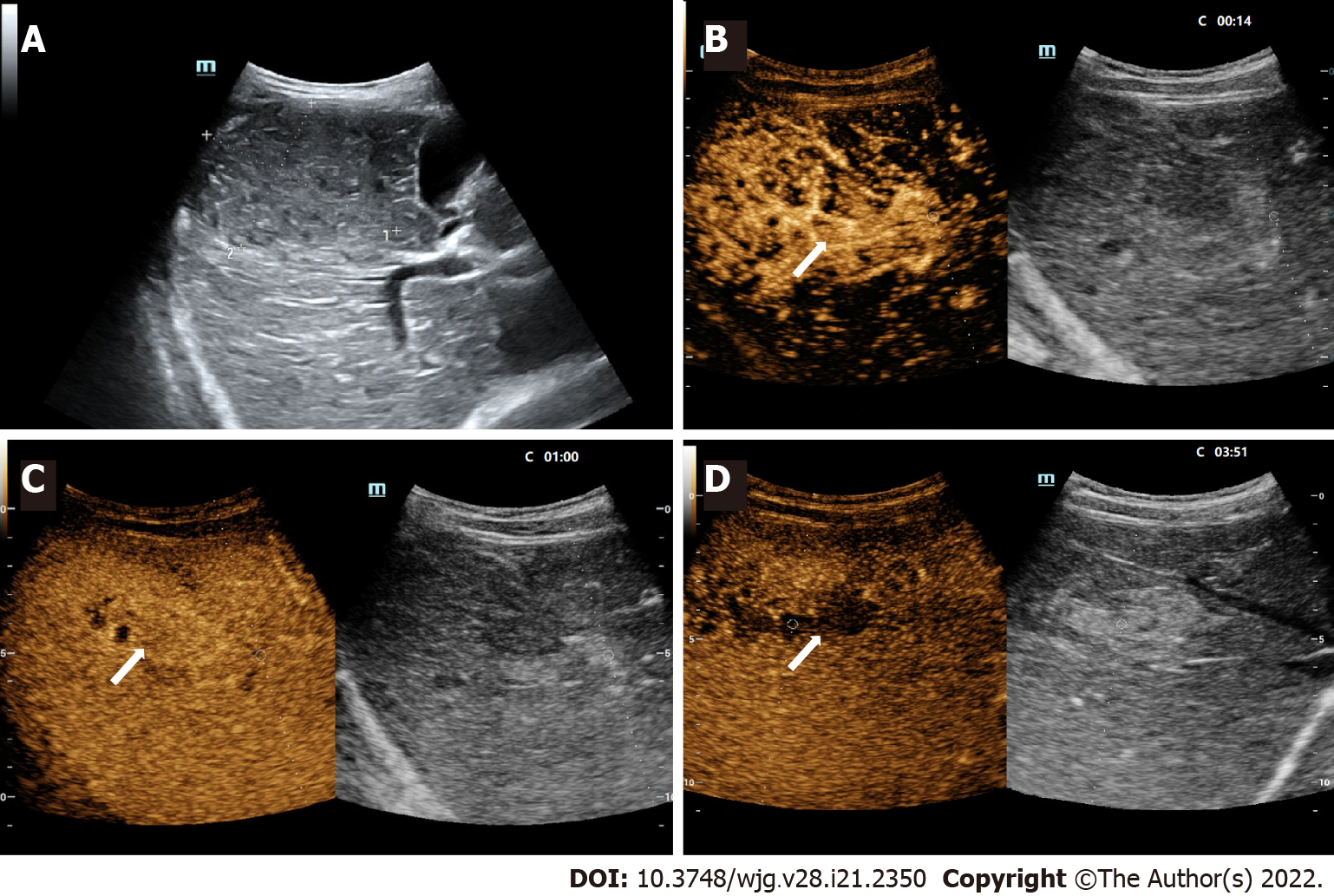
Figure 2 LR-5 nodule in a 10-year-old boy.
A: A hypoechoic nodule (arrow) measuring 7.3 cm in the right lobe of the liver was shown at conventional gray-scale US; B: The lesion was inhomogeneously hyperenhanced (arrow) in the arterial phase (14 s) at contrast-enhanced US; C: The lesion was seen iso-enhanced in the portal phase (60 s); D: Mild washout in the late phase (231 s) was shown. There were small areas of nonenhancement within the lesion during the whole process. The patient had a chronic hepatitis B viral infection. The serum AFP level was greater than 1210 ng/mL. This lesion was assigned to LR-5 and was confirmed as hepatocellular carcinoma by histopathologic analysis.
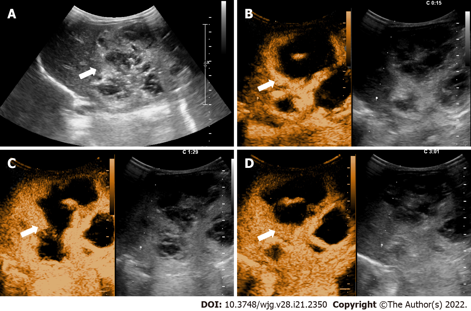
Figure 3 LR-4 nodule in a 19-hour-old newborn.
A: An inhomogeneous hyperechoic nodule measuring 7.2-cm (arrow) in the left lobe of the liver was shown at conventional gray-scale US; B: The lesion was inhomogeneously hyperenhanced (arrow) with large area of unenhancement in the arterial phase (15 s) at contrast-enhanced US; C: The enhanced area of the lesion was seen slightly hyperenhanced (arrow) in the portal phase (89 s); D: The enhanced area of the lesion was seen iso-enhanced (arrow) in the late phase (181 s). There were patchy areas of nonenhancement within the lesion during the whole process. The serum AFP level was greater than 1210 ng/mL. Infantile hemangioendothelioma was confirmed at histopathologic analysis.
- Citation: Jiang ZP, Zeng KY, Huang JY, Yang J, Yang R, Li JW, Qiu TT, Luo Y, Lu Q. Differentiating malignant and benign focal liver lesions in children using CEUS LI-RADS combined with serum alpha-fetoprotein. World J Gastroenterol 2022; 28(21): 2350-2360
- URL: https://www.wjgnet.com/1007-9327/full/v28/i21/2350.htm
- DOI: https://dx.doi.org/10.3748/wjg.v28.i21.2350


