Copyright
©The Author(s) 2022.
World J Gastroenterol. May 28, 2022; 28(20): 2243-2247
Published online May 28, 2022. doi: 10.3748/wjg.v28.i20.2243
Published online May 28, 2022. doi: 10.3748/wjg.v28.i20.2243
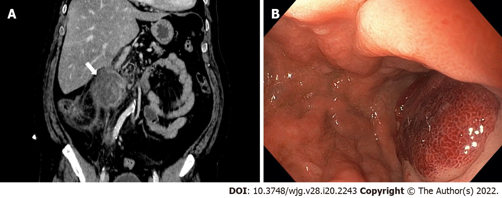
Figure 1 Intramural duodenal hematoma.
A: Contrast-enhanced abdominal computed tomography in coronal section; B: Endoscopic visualization with substenosis of duodenal lumen.
Figure 2 Intramural duodenal hematoma after endoscopic treatment.
A: Endoscopic ultrasonography (EUS) image showed deployment of the distal flange of a cautery-tipped lumen apposing metal stent (LAMS) in the intramural duodenal hematoma under EUS guidance; B: Endoscopic views showed the proximal flange of the LAMS; C: Control abdominal computed tomography confirmed the correct position of the LAMS.
- Citation: Valerii G, Ormando VM, Cellini C, Sacco L, Barbera C. Endoscopic management of intramural spontaneous duodenal hematoma: A case report. World J Gastroenterol 2022; 28(20): 2243-2247
- URL: https://www.wjgnet.com/1007-9327/full/v28/i20/2243.htm
- DOI: https://dx.doi.org/10.3748/wjg.v28.i20.2243


