Copyright
©The Author(s) 2022.
World J Gastroenterol. May 14, 2022; 28(18): 1902-1921
Published online May 14, 2022. doi: 10.3748/wjg.v28.i18.1902
Published online May 14, 2022. doi: 10.3748/wjg.v28.i18.1902
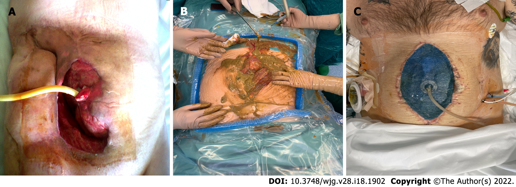
Figure 1 Intra-abdominal sepsis.
A: Abscess in the right iliac fossa, spontaneously draining to the skin, associated with high-flow enteric fistula. Foley catheter placed in the fistulized small intestine; B: Fecal contamination of the peritoneal cavity in a patient with Crohn’s disease with perforation of the transverse colon; C: Open abdomen after treatment of intestinal perforation in Crohn’s disease. Negative pressure intraperitoneal device placement.
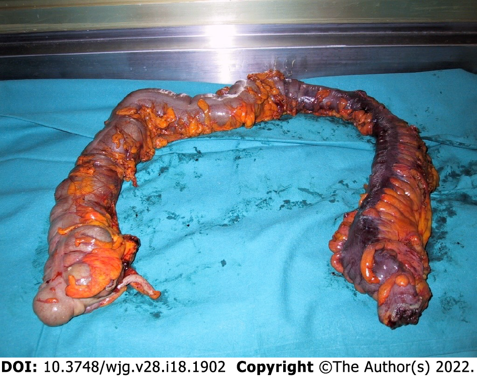
Figure 2 Surgical specimen of total intra-abdominal colectomy in patient with acute colitis.
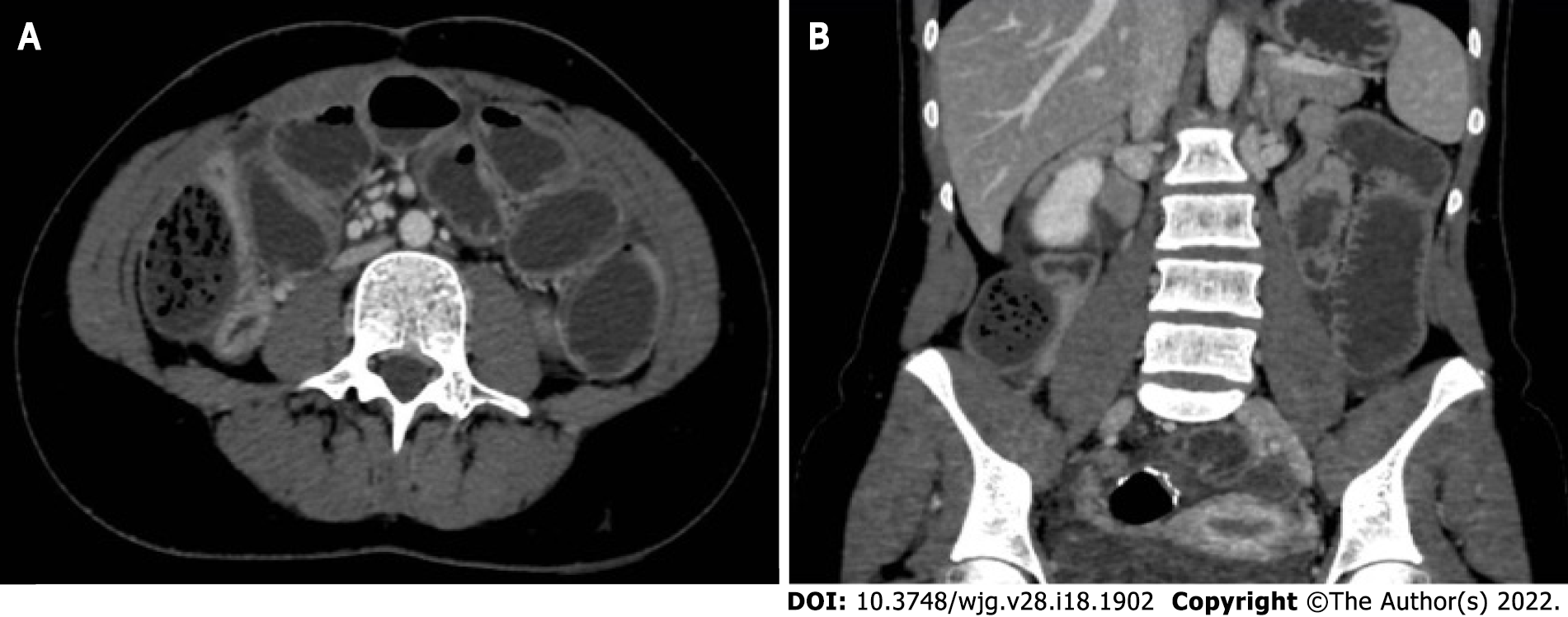
Figure 3 Computed tomography scan.
Coronal view and sagittal view showing postoperative recurrence of Crohn’s disease with small bowel stenosis in patient previously treated by colectomy and ileo-anal anastomosis. A: Coronal view; B: Sagittal view.
- Citation: Chiarello MM, Pepe G, Fico V, Bianchi V, Tropeano G, Altieri G, Brisinda G. Therapeutic strategies in Crohn’s disease in an emergency surgical setting. World J Gastroenterol 2022; 28(18): 1902-1921
- URL: https://www.wjgnet.com/1007-9327/full/v28/i18/1902.htm
- DOI: https://dx.doi.org/10.3748/wjg.v28.i18.1902


