Copyright
©The Author(s) 2022.
World J Gastroenterol. May 7, 2022; 28(17): 1860-1870
Published online May 7, 2022. doi: 10.3748/wjg.v28.i17.1860
Published online May 7, 2022. doi: 10.3748/wjg.v28.i17.1860
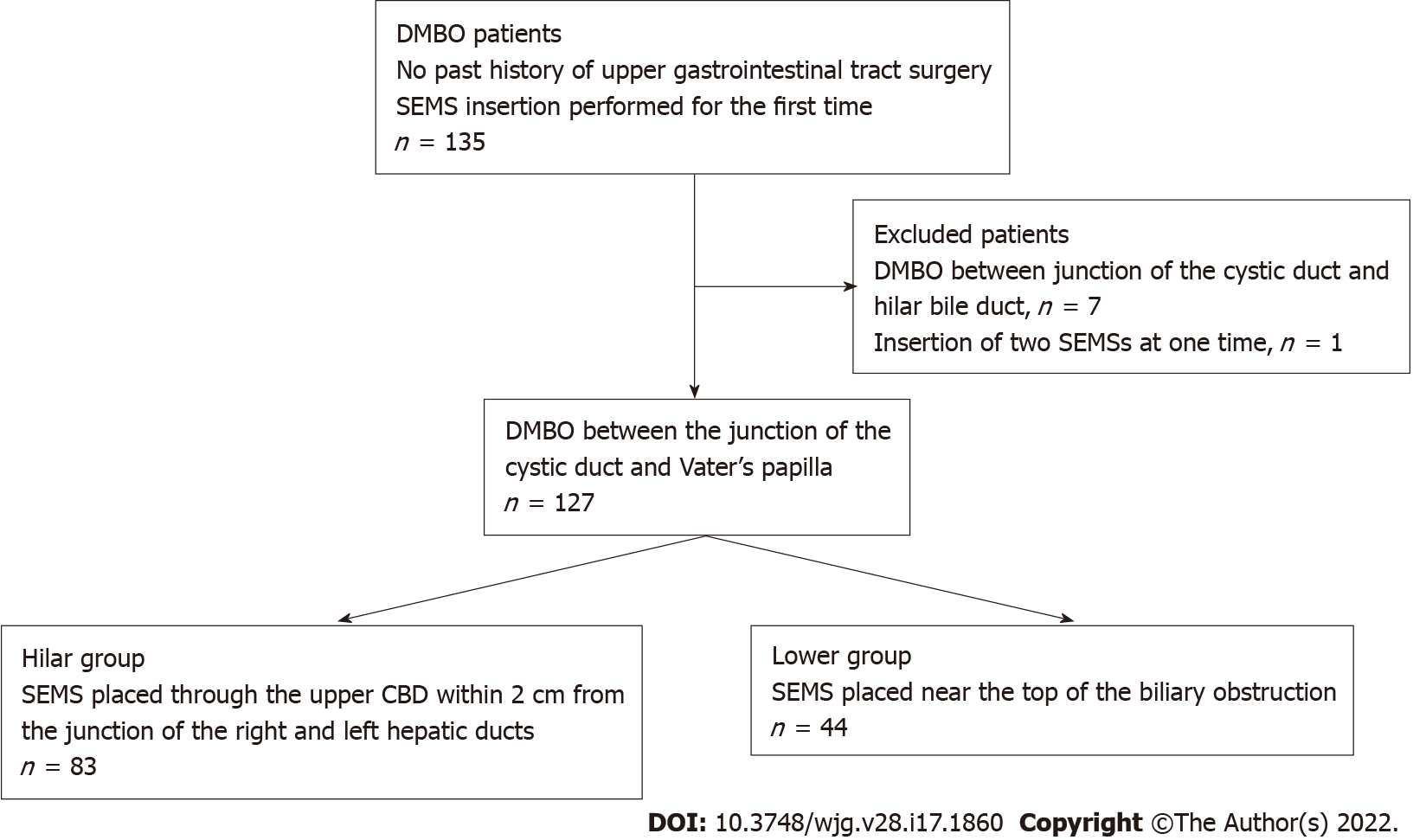
Figure 1 Patient flowchart.
DMBO: Distal malignant biliary obstruction; SEMS: Self-expandable metallic stent; CBD: Common bile duct.
Figure 2 Representative cases from each group.
A and B: A patient with distal malignant biliary obstruction (DMBO) in the Hilar group who underwent self-expandable metallic stent (SEMS) placement near the biliary hilar duct; C and D: A patient with DMBO in the Lower group who underwent SEMS placement near the top of the biliary obstruction.
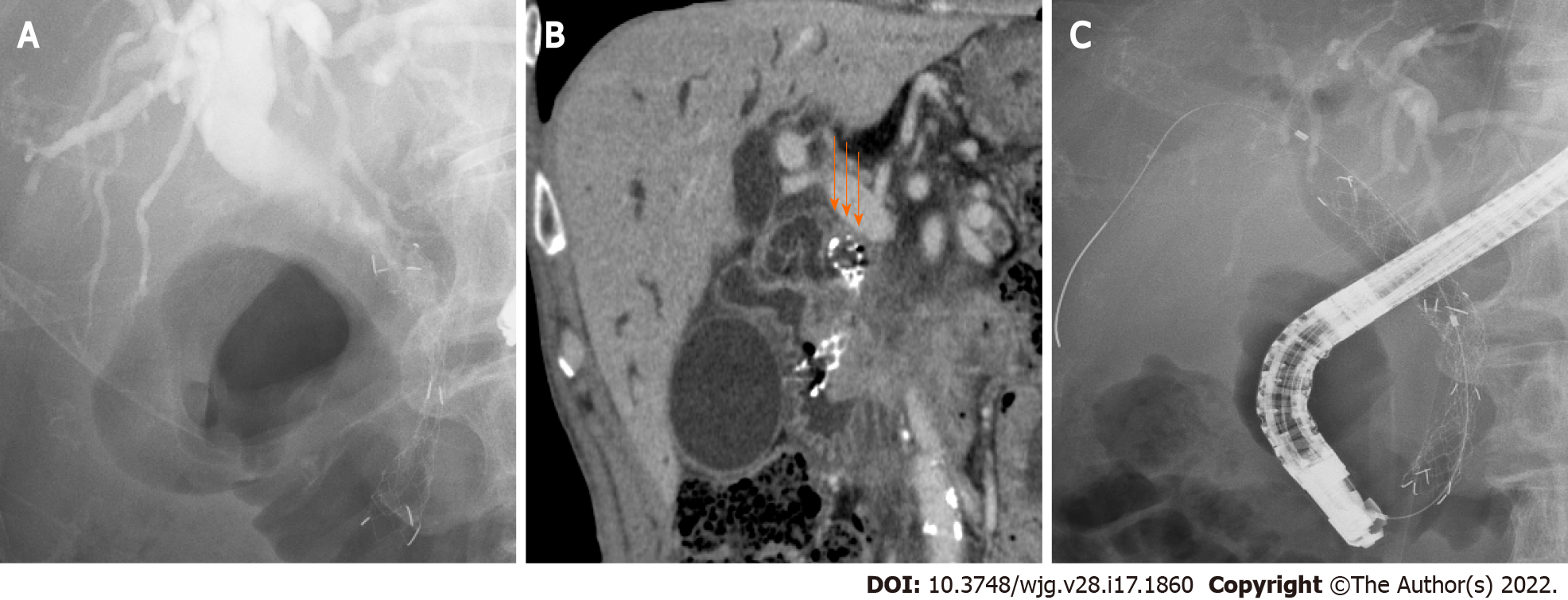
Figure 3 A patient with closure of the top edge of the self-expandable metallic stent by the common bile duct wall.
A: A patient with distal malignant biliary obstruction who underwent uncovered self-expandable metallic stents (USEMS) placement near the top of the biliary obstruction; B: The top edge of the SEMS was closed by the common bile duct wall (arrows). Upper bile tract dilation was observed; C: An additional USEMS was placed near the biliary hilar duct.
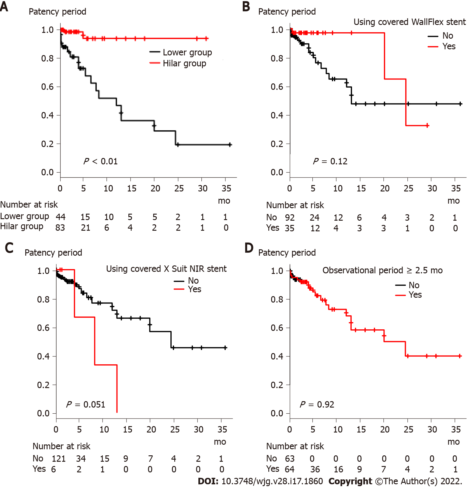
Figure 4 Comparison of stent patency period based on factors that were significantly different between the Hilar group and Lower group.
A: Stent placement position (Hilar group vs Lower group); B: Use of the covered WallFlex stent; C: Use of the covered X Suit NIR stent; D: Observational period (< 2.5 mo vs ≥ 2.5 mo).
- Citation: Sugimoto M, Takagi T, Suzuki R, Konno N, Asama H, Sato Y, Irie H, Okubo Y, Nakamura J, Takasumi M, Hashimoto M, Kato T, Kobashi R, Yanagita T, Hikichi T, Ohira H. Biliary metal stents should be placed near the hilar duct in distal malignant biliary stricture patients. World J Gastroenterol 2022; 28(17): 1860-1870
- URL: https://www.wjgnet.com/1007-9327/full/v28/i17/1860.htm
- DOI: https://dx.doi.org/10.3748/wjg.v28.i17.1860


