Copyright
©The Author(s) 2022.
World J Gastroenterol. Apr 28, 2022; 28(16): 1608-1624
Published online Apr 28, 2022. doi: 10.3748/wjg.v28.i16.1608
Published online Apr 28, 2022. doi: 10.3748/wjg.v28.i16.1608
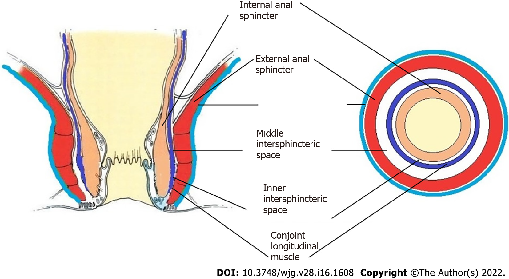
Figure 1 Normal anatomy.
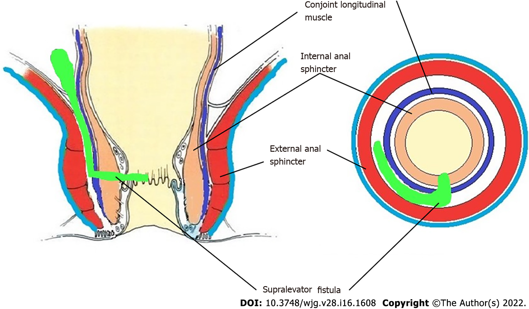
Figure 2 Supralevator fistula.
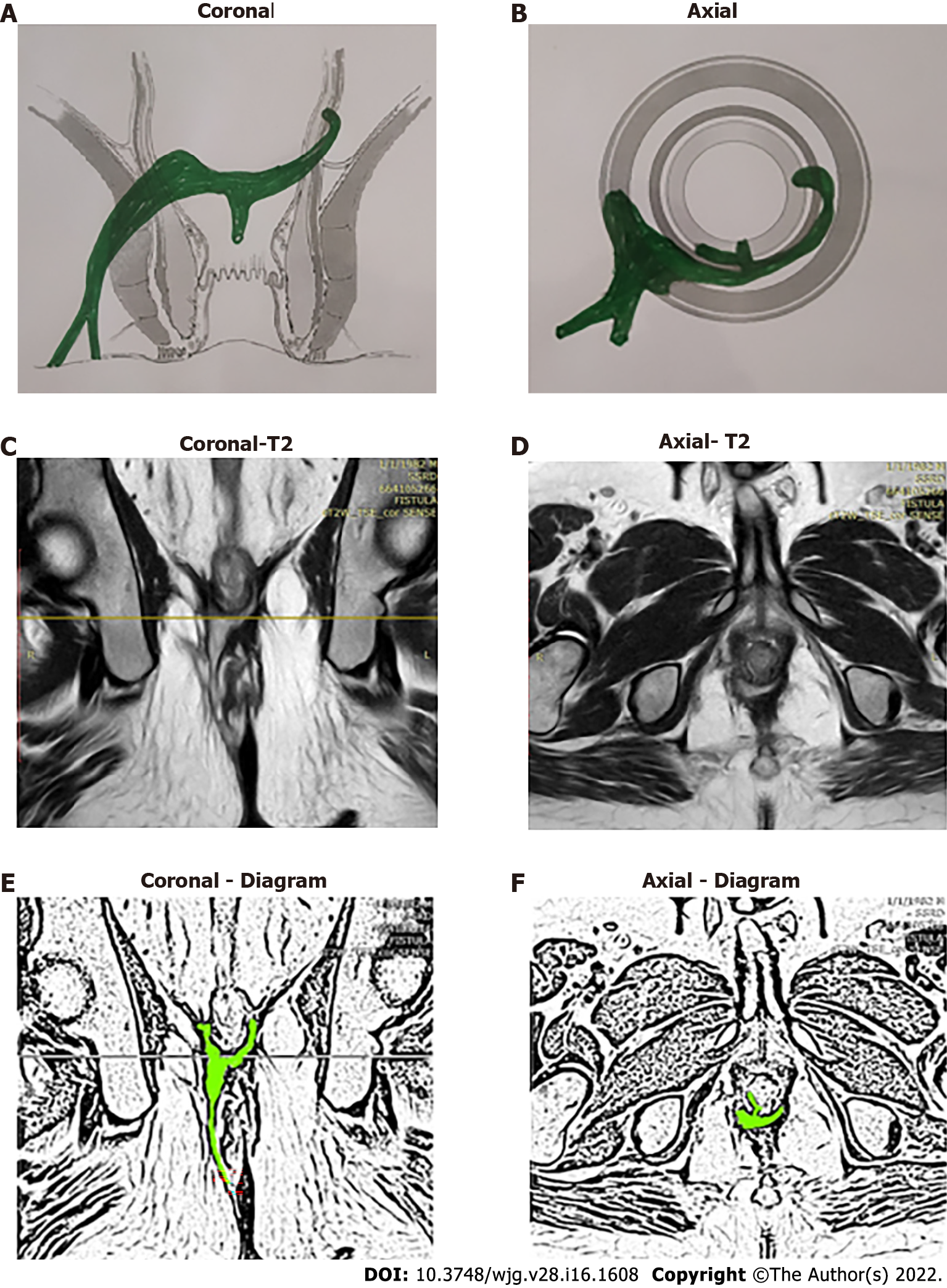
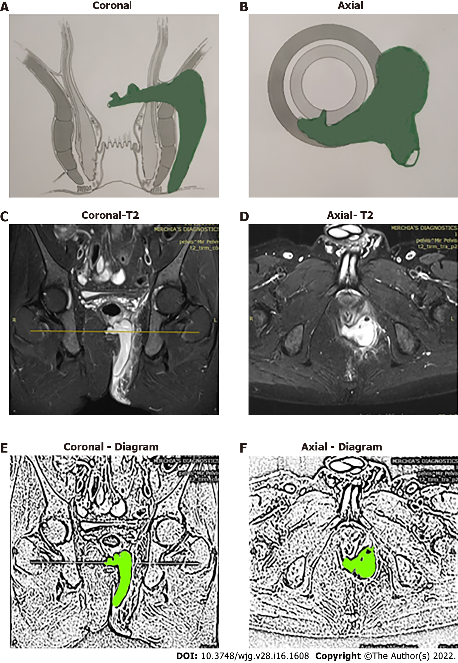
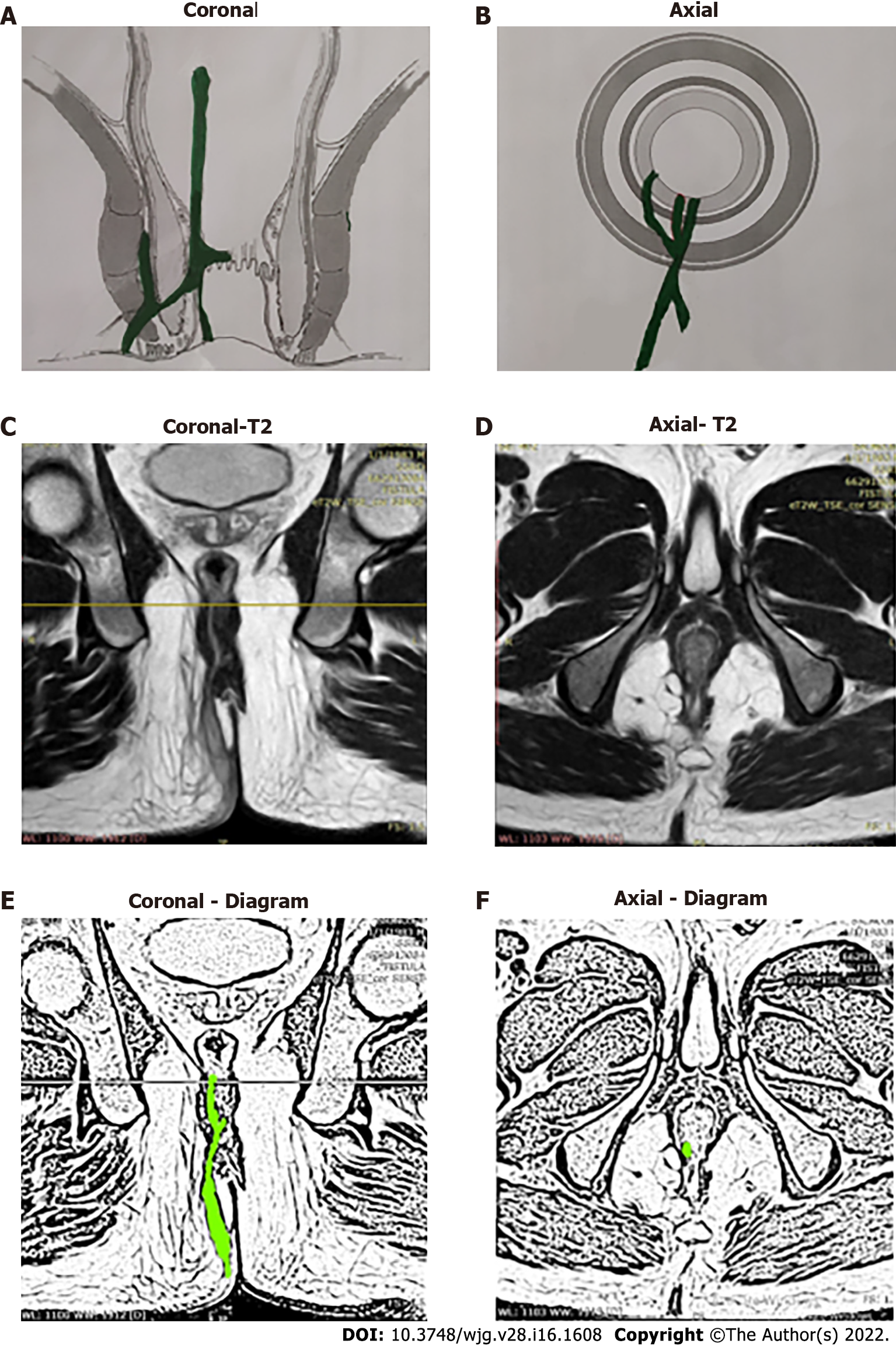
Figure 3 A 39-year-old male patient with a high transsphincteric fistula with supralevator extension at 3 o’clock.
There is a high intrarectal tract from 6 to 7 o’clock. The internal opening is at 6 o’clock position. A: Coronal section (schematic diagram); B: Axial section (schematic diagram); C: T2-weighted magnetic resonance imaging (MRI) Coronal section showing supralevator tract; D: T2-weighted MRI Axial section; E: Sketch of MRI Coronal section highlighting fistula tract (light green color); F: Sketch of MRI Axial section highlighting fistula tract (light green color).
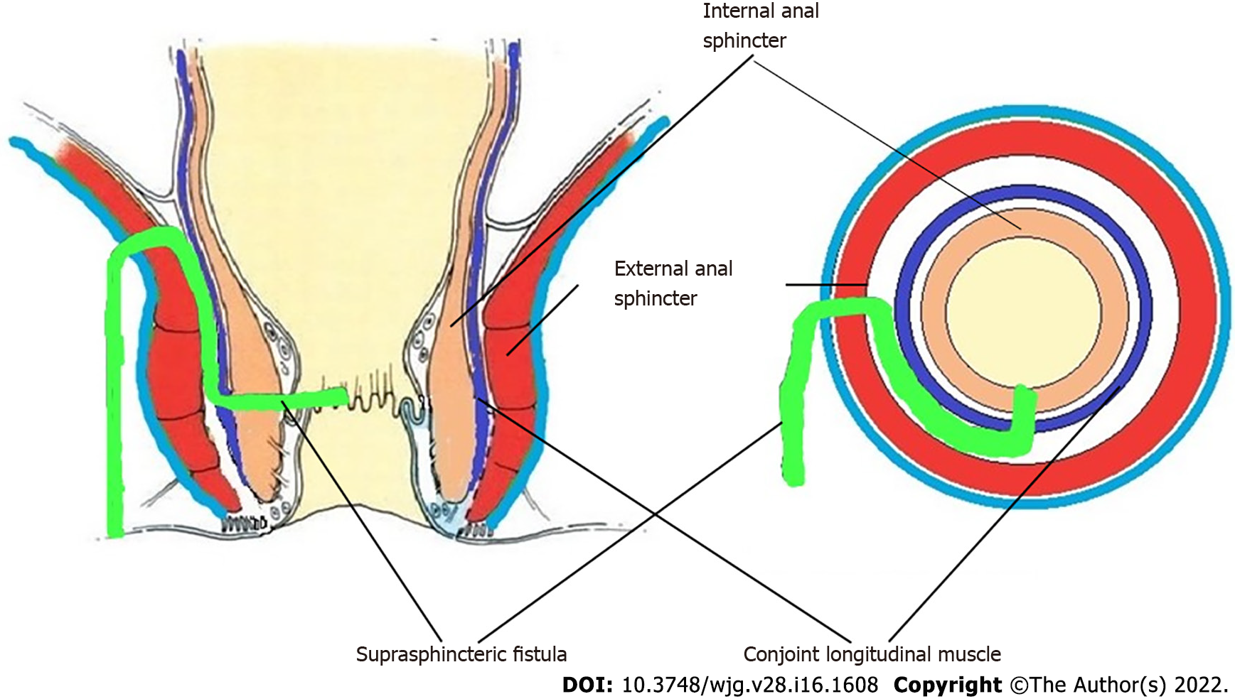
Figure 4 Suprasphincteric fistula.
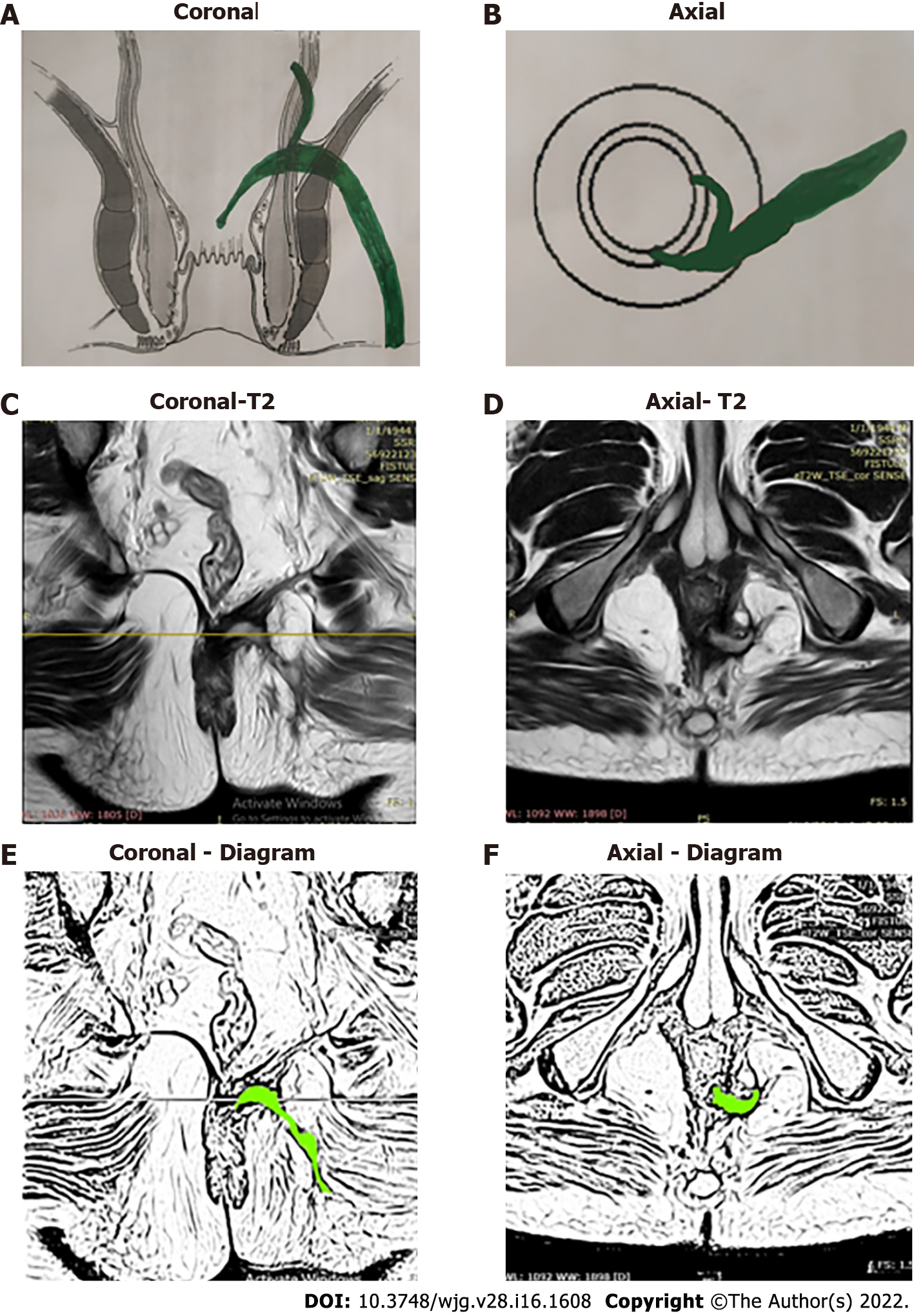
Figure 5 A 40-year-old male patient with a suprasphincteric fistula and abscess on left side.
The internal opening is at 6:30 o’clock position. A: Coronal section (schematic diagram); B: Axial section (schematic diagram); C: Short Tau Inversion Recovery (STIR) magnetic resonance imaging (MRI) Coronal section showing suprasphincteric tract and abscess; D: STIR MRI Axial section; E: Sketch of MRI Coronal section highlighting suprasphincteric fistula tract and abscess (light green color); F: Sketch of MRI Axial section highlighting fistula tract and abscess (light green color).
Figure 6 A 47-year-old male patient with a suprasphincteric fistula on left side and supralevator tract and high rectal opening at 3 o’clock.
The internal opening is at 6 o’clock position. A: Coronal section (schematic diagram); B: Axial section (schematic diagram); C: T2-weighted magnetic resonance imaging (MRI) Coronal section showing suprasphincteric tract; D: T2-weighted MRI Axial section; E: Sketch of MRI Coronal section highlighting supralevator fistula tract (light green color); F: Sketch of MRI Axial section highlighting fistula tract (light green color).
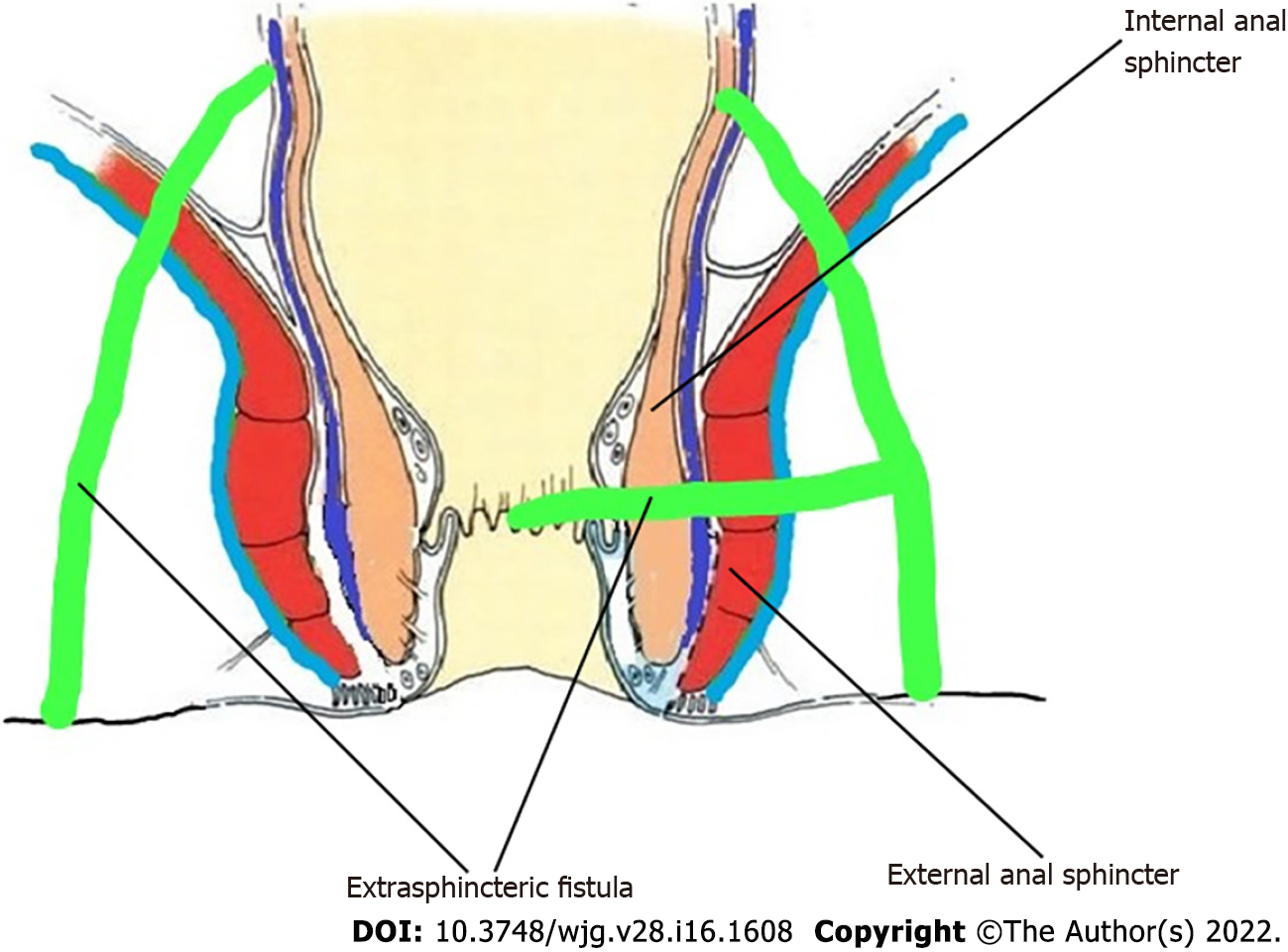
Figure 7 Extrasphincteric fistula.
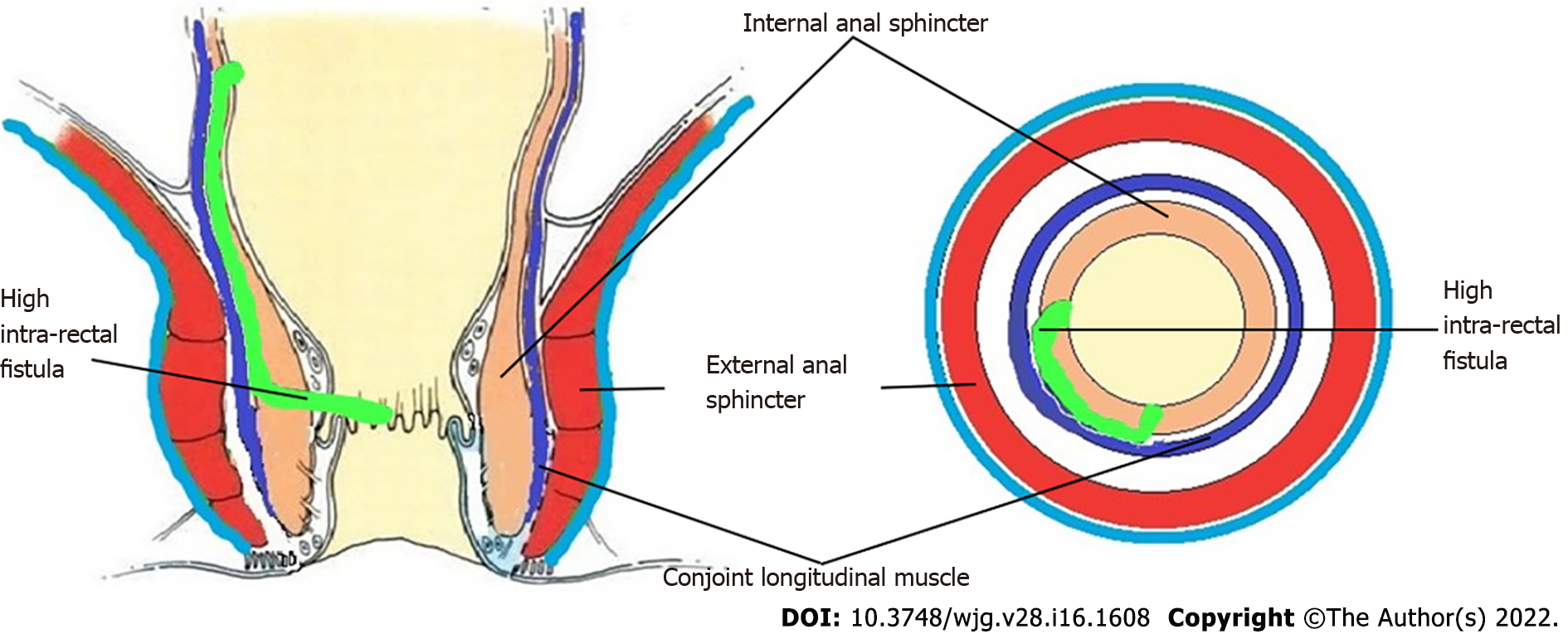
Figure 8 High Intrarectal fistula.
Figure 9 A 36-year-old male patient with a high intrarectal fistula at 7 o’clock.
A: Coronal section (schematic diagram); B: Axial section (schematic diagram); C: T2-weighted magnetic resonance imaging (MRI) Coronal section showing high intrarectal fistula tract; D: T2-weighted MRI Axial section; E: Sketch of MRI Coronal section highlighting high intrarectal fistula tract (light green color); F: Sketch of MRI Axial section highlighting fistula tract (light green color).
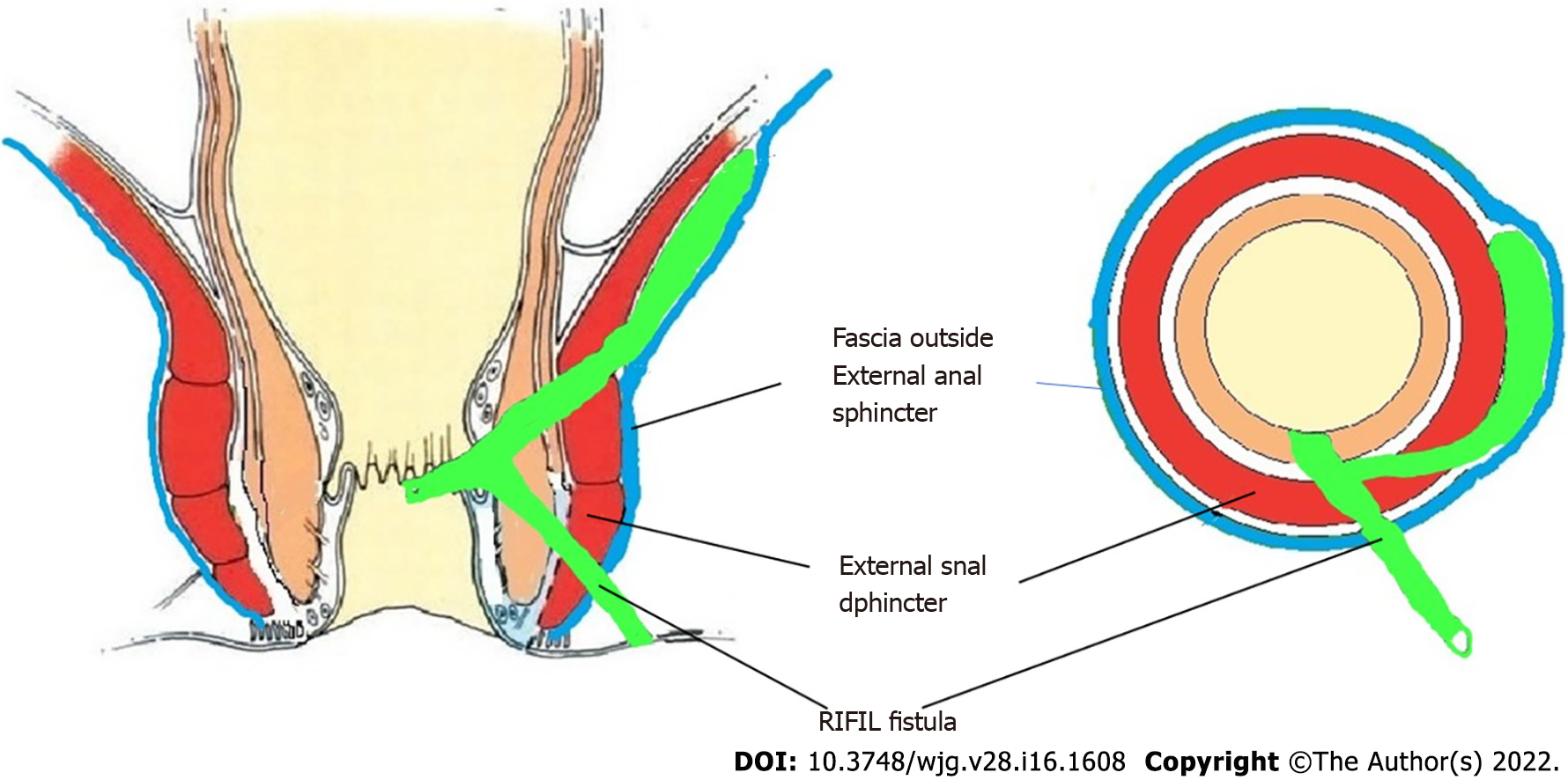
Figure 10 Roof of ischiorectal fossa inside the levator ani muscle fistula.
RIFIL: Roof of ischiorectal fossa inside the levator ani muscle.
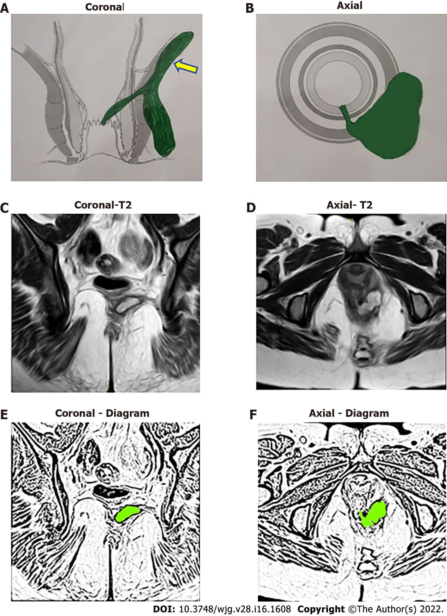
Figure 11 A 33-year-old female patient with a roof of ischiorectal fossa inside the levator ani muscle fistula on left side at 5 o’clock and internal opening at 6 o’clock position.
A: Coronal section showing roof of ischiorectal fossa inside the levator ani muscle (RIFIL) fistula (yellow arrow) (schematic diagram); B: Axial section (schematic diagram); C: T2-weighted magnetic resonance imaging (MRI) Coronal section showing RIFIL fistula tract; D: T2-weighted MRI Axial section; E: Sketch of MRI Coronal section highlighting RIFIL fistula tract (light green color); F: Sketch of MRI Axial section highlighting fistula tract (light green color).
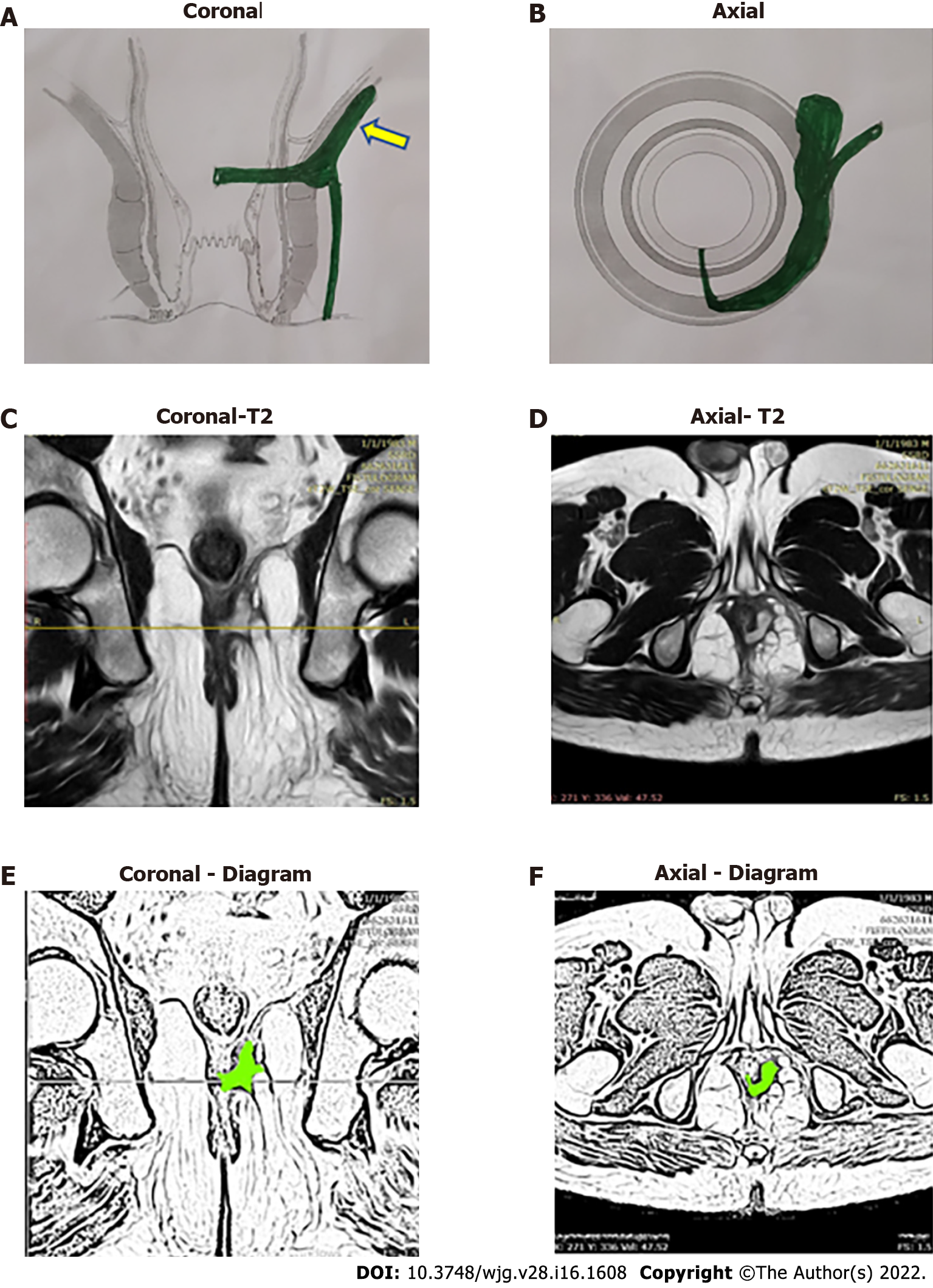
Figure 12 A 38-year-old male patient with a roof of ischiorectal fossa inside the levator ani muscle fistula on left side at 3 o’clock and internal opening at 6 o’clock position.
A: Coronal section showing roof of ischiorectal fossa inside the levator ani muscle (RIFIL) fistula (yellow arrow) (schematic diagram); B: Axial section (schematic diagram); C: T2-weighted magnetic resonance imaging (MRI) Coronal section showing RIFIL fistula tract; D: T2-weighted MRI Axial section; E: Sketch of MRI Coronal section highlighting RIFIL fistula tract (light green color); F: Sketch of MRI Axial section highlighting fistula tract (light green color).
- Citation: Garg P, Yagnik VD, Dawka S, Kaur B, Menon GR. Guidelines to diagnose and treat peri-levator high-5 anal fistulas: Supralevator, suprasphincteric, extrasphincteric, high outersphincteric, and high intrarectal fistulas. World J Gastroenterol 2022; 28(16): 1608-1624
- URL: https://www.wjgnet.com/1007-9327/full/v28/i16/1608.htm
- DOI: https://dx.doi.org/10.3748/wjg.v28.i16.1608


