Copyright
©The Author(s) 2022.
World J Gastroenterol. Apr 7, 2022; 28(13): 1304-1314
Published online Apr 7, 2022. doi: 10.3748/wjg.v28.i13.1304
Published online Apr 7, 2022. doi: 10.3748/wjg.v28.i13.1304
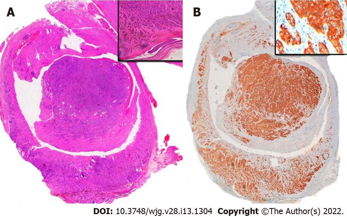
Figure 1 Histological images.
A: Well-differentiated aNET that infiltrate the entire wall of the appendix and focally infiltrate the adjacent fat, affecting the surgical margin; B: Immunohistochemical techniques reveal positivity for synaptophysin and CgA.
- Citation: Muñoz de Nova JL, Hernando J, Sampedro Núñez M, Vázquez Benítez GT, Triviño Ibáñez EM, del Olmo García MI, Barriuso J, Capdevila J, Martín-Pérez E. Management of incidentally discovered appendiceal neuroendocrine tumors after an appendicectomy. World J Gastroenterol 2022; 28(13): 1304-1314
- URL: https://www.wjgnet.com/1007-9327/full/v28/i13/1304.htm
- DOI: https://dx.doi.org/10.3748/wjg.v28.i13.1304


