Copyright
©The Author(s) 2022.
World J Gastroenterol. Mar 21, 2022; 28(11): 1139-1158
Published online Mar 21, 2022. doi: 10.3748/wjg.v28.i11.1139
Published online Mar 21, 2022. doi: 10.3748/wjg.v28.i11.1139
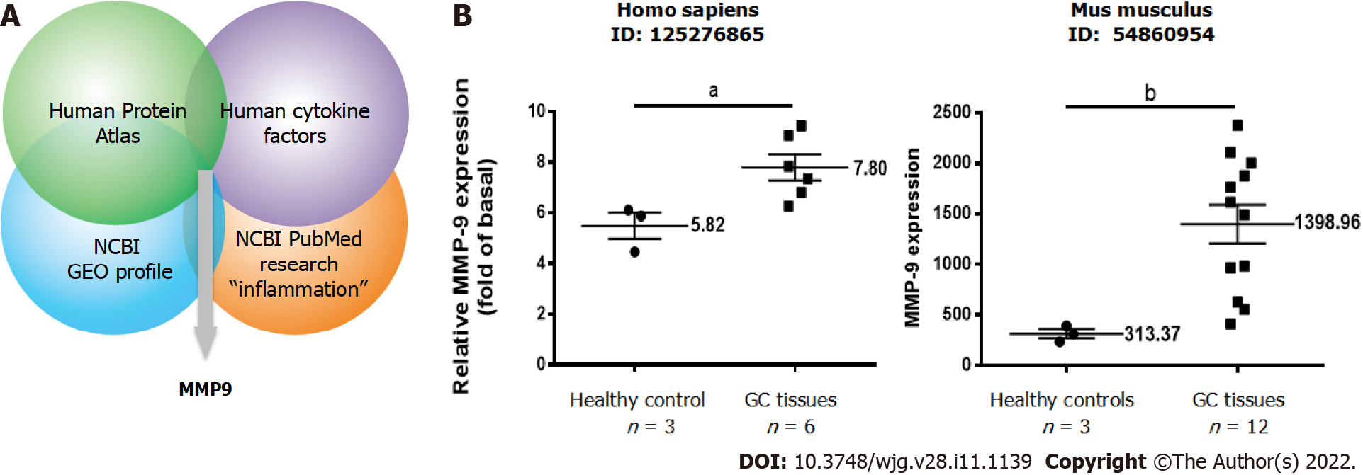
Figure 1 Identification studies of matrix metallopeptidase-9, a potential inflammation-related protein involved in gastric cancer.
A: Identification of potential inflammation-related proteins involved in gastric cancer (GC) based on an omics approach including four integrated datasets. The strategy included proteomic profiling; B: Relative expression levels of matrix metallopeptidase-9 in paired GC and adjacent normal tissues in humans and mice. A t test was used for comparisons between the two groups. Data are expressed as the mean ± SEM of three independent experiments. aP < 0.05, bP < 0.01 vs controls. GC: Gastric cancer; GEO: Gene Expression Omnibus; MMP-9: Matrix metallopeptidase-9; NCBI: National Center for Biotechnology Information.
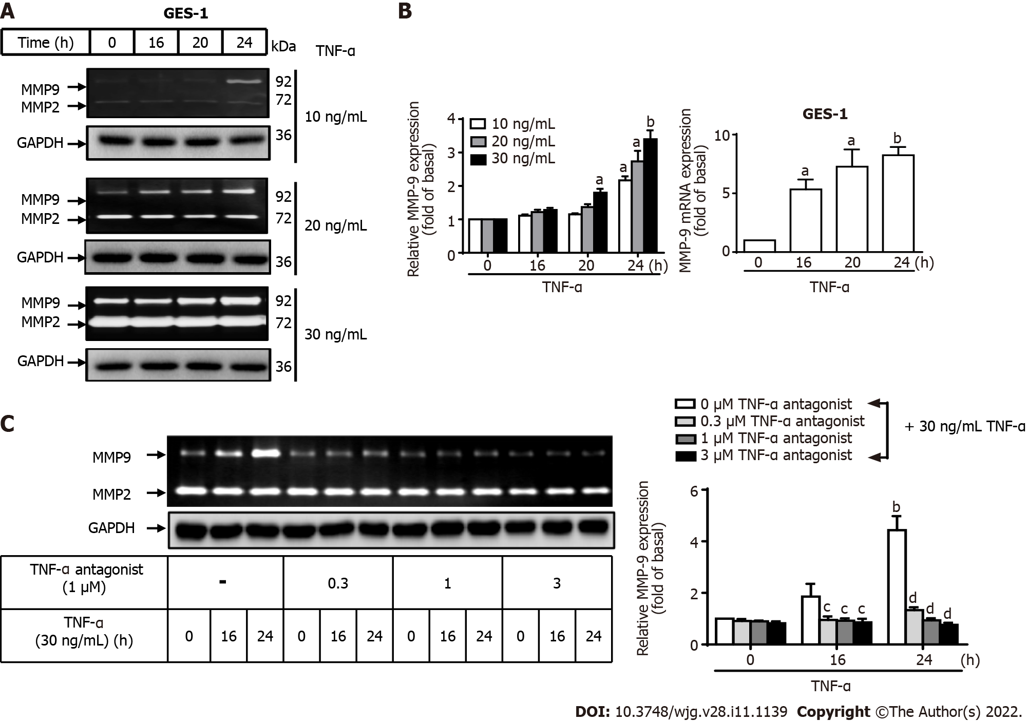
Figure 2 Tumor necrosis factor-α induces matrix metallopeptidase-9 expression via tumor necrosis factor-α receptor in normal human gastric mucosa epithelial cells.
A: Tumor necrosis factor-α (TNF-α) was used at concentrations of 10, 20, and 30 ng/mL to stimulate normal human gastric mucosa epithelial cells (GES-1) for 0, 16, 20, or 24 h, and matrix metallopeptidase-9 (MMP-9) enzymatic activity was measured by gelatin zymography as described in the Materials and Methods section; B: MMP-9 transcripts were analyzed by quantitative reverse transcription-polymerase chain reaction; C: Cells were either untreated or treated with a TNF-α antagonist (TNFR inhibitor) (1 mM) 1 h before the addition of TNF-α (30 ng/mL). The TNF-α antagonist repressed TNF-α-activated MMP-9 expression in GES-1 cells. One-way ANOVA was used for comparisons among different treatment time points (aP < 0.05, bP < 0.01 vs control cells in 0 h). One-way ANOVA was used for comparisons among different treatments (cP < 0.05, dP < 0.01 vs TNF-α-stimulated cells). Data are expressed as the mean ± SEM of three independent experiments. GAPDH: Glyceraldehyde-3-phosphate dehydrogenase; GES-1: Normal human gastric mucosa epithelial cell line; MMP: Matrix metallopeptidase; TNF-α: Tumor necrosis factor-α.
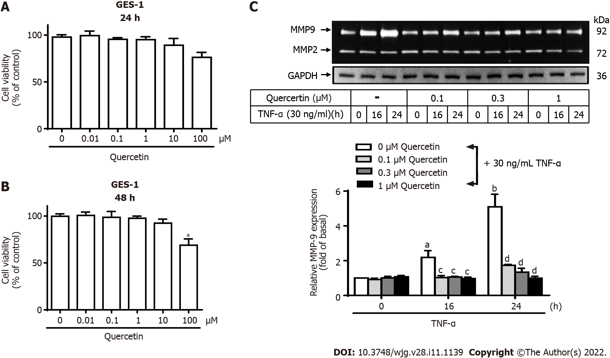
Figure 3 Quercetin suppresses tumor necrosis factor-α-induced matrix metallopeptidase-9 expression in normal human gastric mucosa epithelial cells.
A and B: Effects of quercetin on the viability of normal human gastric mucosa epithelial cells (GES-1). GES-1 cells were treated with different concentrations of quercetin (0, 0.01, 0.1, 1, 10, or 100 mM) for 24 h (A) or 48 h (B), and cell viability was measured using the cell counting Kit-8 assay; C: Cells were either untreated or treated with quercetin (0, 0.1, 0.3, or 1 mM) for 1 h before the addition of tumor necrosis factor-α (TNF-α) (30 ng/mL) and then incubated for 0, 16, or 24 h as described in the Materials and Methods section. Matrix metallopeptidase-9 zymogen activity was analyzed by gelatin zymography. One-way ANOVA was used for comparisons among different treatment time points (aP < 0.05, bP < 0.01 vs control cells in 0 h). One-way ANOVA was used for comparisons among different treatments (cP < 0.05, dP < 0.01 vs TNF-α-stimulated cells). Data are expressed as the mean ± SEM of three independent experiments. CCK-8: Cell Counting Kit-8; GAPDH: Glyceraldehyde-3-phosphate dehydrogenase; GES-1: Normal human gastric mucosa epithelial cell line; MMP-9: Matrix metallopeptidase-9; TNF-α: Tumor necrosis factor-α.
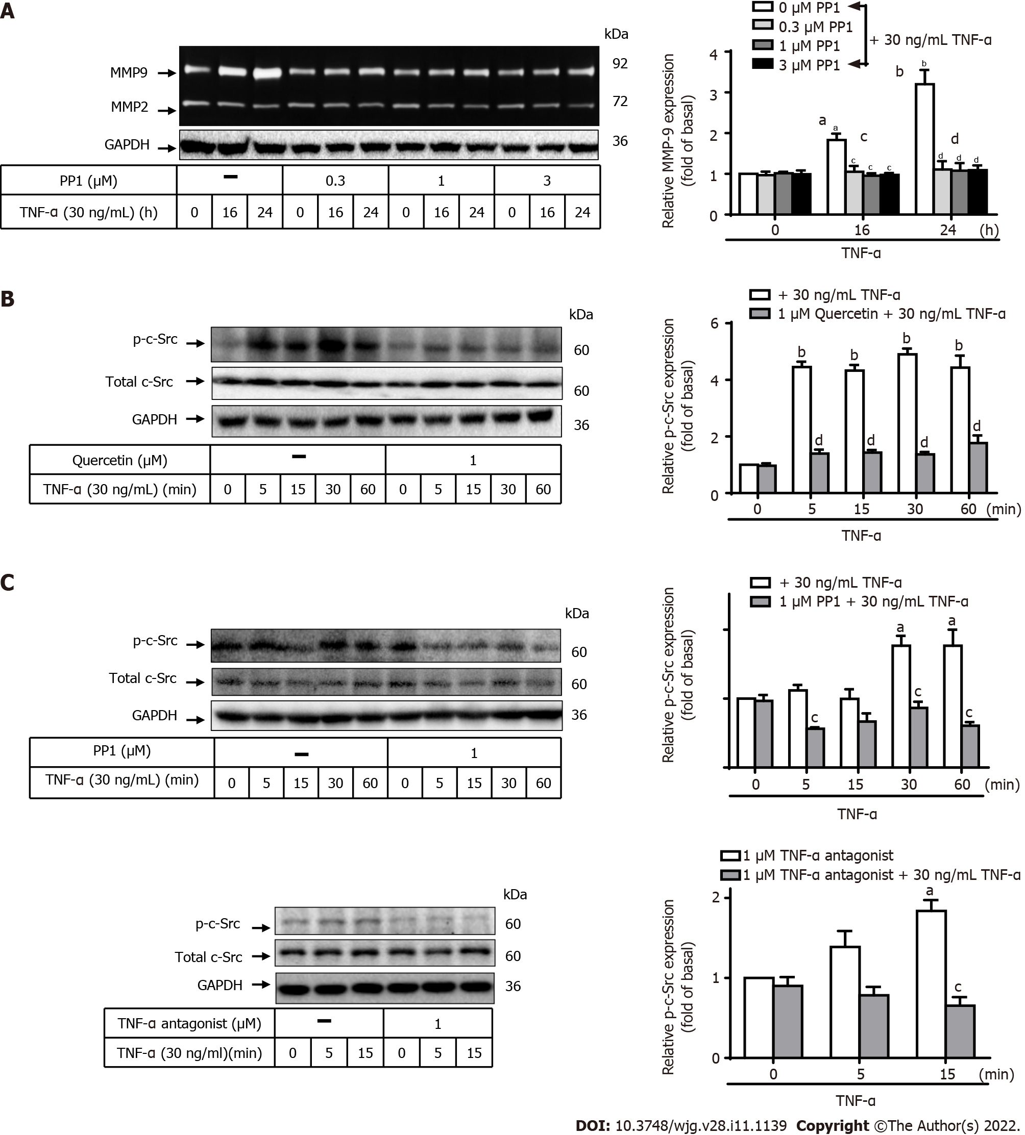
Figure 4 Quercetin suppresses tumor necrosis factor-α-induced matrix metallopeptidase-9 expression via tumor necrosis factor-α antagonist-proto-oncogene tyrosine-protein kinase Src in normal human gastric mucosa epithelial cells.
A: Cells were either untreated or treated with PP1 (0, 0.3, 1, or 3 mM) for 1 h before the addition of tumor necrosis factor-α (TNF-α) (30 ng/mL) and then incubated for 0, 16, or 24 h as described in the Materials and Methods section. Matrix metallopeptidase-9 (MMP-9) zymogen activity was analyzed by gelatin zymography; B and C: Cells were either untreated or treated with quercetin (B; 0 or 1 mM) or PP1 (C; 0 or 1 mM) or TNF-α antagonist (1 mM) for 1 h before the addition of TNF-α (30 ng/mL) and incubated for 0, 16, or 24 h. The phosphorylation of c-Src was measured using Western blot analysis after incubation of cells with TNF-α for 0, 5, 15, 30, or 60 min. One-way ANOVA was used for comparisons among different treatment time points (aP < 0.05, bP < 0.01 vs control cells in 0 h). One-way ANOVA was used for comparisons among different treatments (cP < 0.05, dP < 0.01 vs TNF-α-stimulated cells). Data are expressed as the mean ± SEM of three independent experiments. c-Src: Proto-oncogene tyrosine-protein kinase Src; GAPDH: Glyceraldehyde-3-phosphate dehydrogenase; GES-1: Normal human gastric mucosa epithelial cell line; MMP-9: Matrix metallopeptidase-9; TNF-α: Tumor necrosis factor-α.
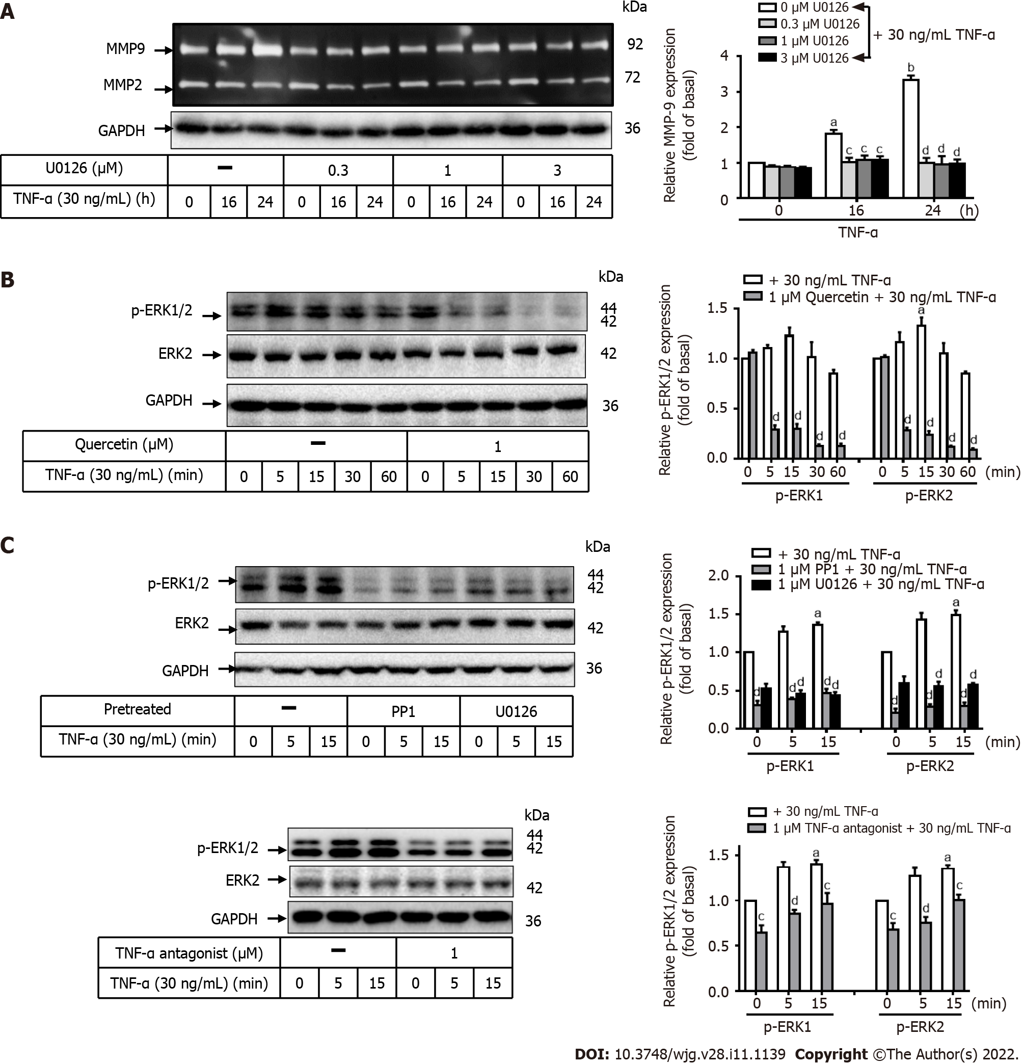
Figure 5 Quercetin suppresses tumor necrosis factor-α-induced matrix metallopeptidase-9 expression via tumor necrosis factor-α antagonist-c-Src-extracellular-signal-regulated kinase 1/2 in normal human gastric mucosa epithelial cells.
A: Normal human gastric mucosa epithelial cells (GES-1) were either untreated or treated with U0126 (0, 0.3, 1, or 3 mM) for 1 h before the addition of tumor necrosis factor-α (TNF-α) (30 ng/mL) and incubated for 0, 16, or 24 h as described in the Materials and Methods section. Matrix metallopeptidase-9 zymogen activity was measured using gelatin zymography; B and C: Cells were either untreated or treated with quercetin (B; 0 or 1 mM), PP1 (C; 0 or 1 mM), U0126 (0 or 1 mM), or TNF-α antagonist (1 mM) for 1 h before the addition of TNF-α (30 ng/mL) and incubated for 0, 16, or 24 h. The phosphorylation of extracellular-signal-regulated kinase 1/2 was examined by Western blot analysis in cells incubated for 0, 5, 15, 30, or 60 min. One-way ANOVA was used for comparisons among different treatment time points (aP < 0.05, bP < 0.01 vs control cells in 0 h). One-way ANOVA was used for comparisons among different treatments (cP < 0.05, dP < 0.01 vs TNF-α-stimulated cells). Data are expressed as the mean ± SEM of three independent experiments. ERK: Extracellular-signal-regulated kinase; GAPDH: Glyceraldehyde-3-phosphate dehydrogenase; GC: Gastric cancer; GES-1: Normal human gastric mucosa epithelial cell line; MMP-9: Matrix metallopeptidase-9; TNF-α: Tumor necrosis factor-α.
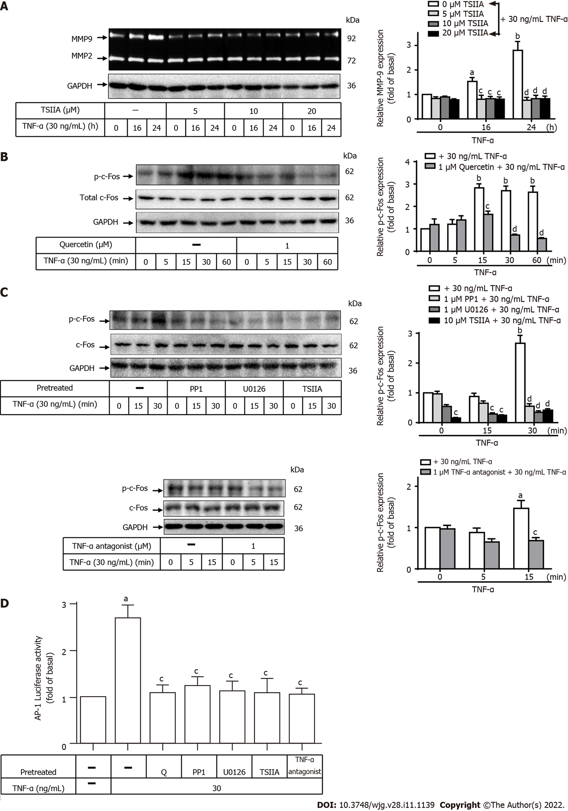
Figure 6 Quercetin suppresses tumor necrosis factor-α-induced matrix metallopeptidase-9 expression via tumor necrosis factor-α antagonist-c-Src-extracellular-signal-regulated kinase 1/2–c-Fos in normal human gastric mucosa epithelial cells.
A: Normal human gastric mucosa epithelial cells (GES-1) were either untreated or treated with Tanshinone IIA (TSIIA) (0, 5, 10, or 20 mM) 1 h before the addition of tumor necrosis factor-α (TNF-α) (30 ng/mL) and incubated for 0, 16, or 24 h as described in the Materials and Methods section. Matrix metallopeptidase-9 zymogen activity was measured using gelatin zymography; B and C: Cells were either untreated or treated with quercetin (B; 0 or 1 mM), PP1 (C; 0 or 1 mM), U0126 (0 or 1 mM), TSIIA (0 or 10 mM), or TNF-α antagonist (1 mM) for 1 h before the addition of TNF-α (30 ng/mL) and incubated for 0, 16, or 24 h. The phosphorylation of c-Fos was measured by Western blot analysis in cells incubated for 0, 5, 15, 30, or 60 min; D: GES-1 cells were transfected with human AP-1–Luc response element reporter plasmids containing the dual-luciferase reporter system. The cells were then pretreated with PP1 (0 or 1 mM), U0126 (0 or 1 mM), TSIIA (0 or 10 mM), TNF-α antagonist (1 mM), or quercetin (1 mM) for 1 h and exposed to TNF-α for 1 h, and luciferase activity was measured. Firefly luciferase activity was standardized to Renilla luciferase activity. One-way ANOVA was used for comparisons among different treatment time points (aP < 0.05, bP < 0.01 vs control cells in 0 h). One-way ANOVA was used for comparisons among different treatments (cP < 0.05, dP < 0.01 vs TNF-α-stimulated cells). Data are expressed as the mean ± SEM of three independent experiments. ERK: Extracellular-signal-regulated kinase; AP-1: Activator protein-1; GAPDH: Glyceraldehyde-3-phosphate dehydrogenase; GES-1: Normal human gastric mucosa epithelial cell line; Luc: Luciferase; GES-1: Normal human gastric mucosa epithelial cell line; MMP-9: Matrix metallopeptidase-9; Q: quercetin; TNF-α: Tumor necrosis factor-α; TSIIA: Tanshinone IIA.
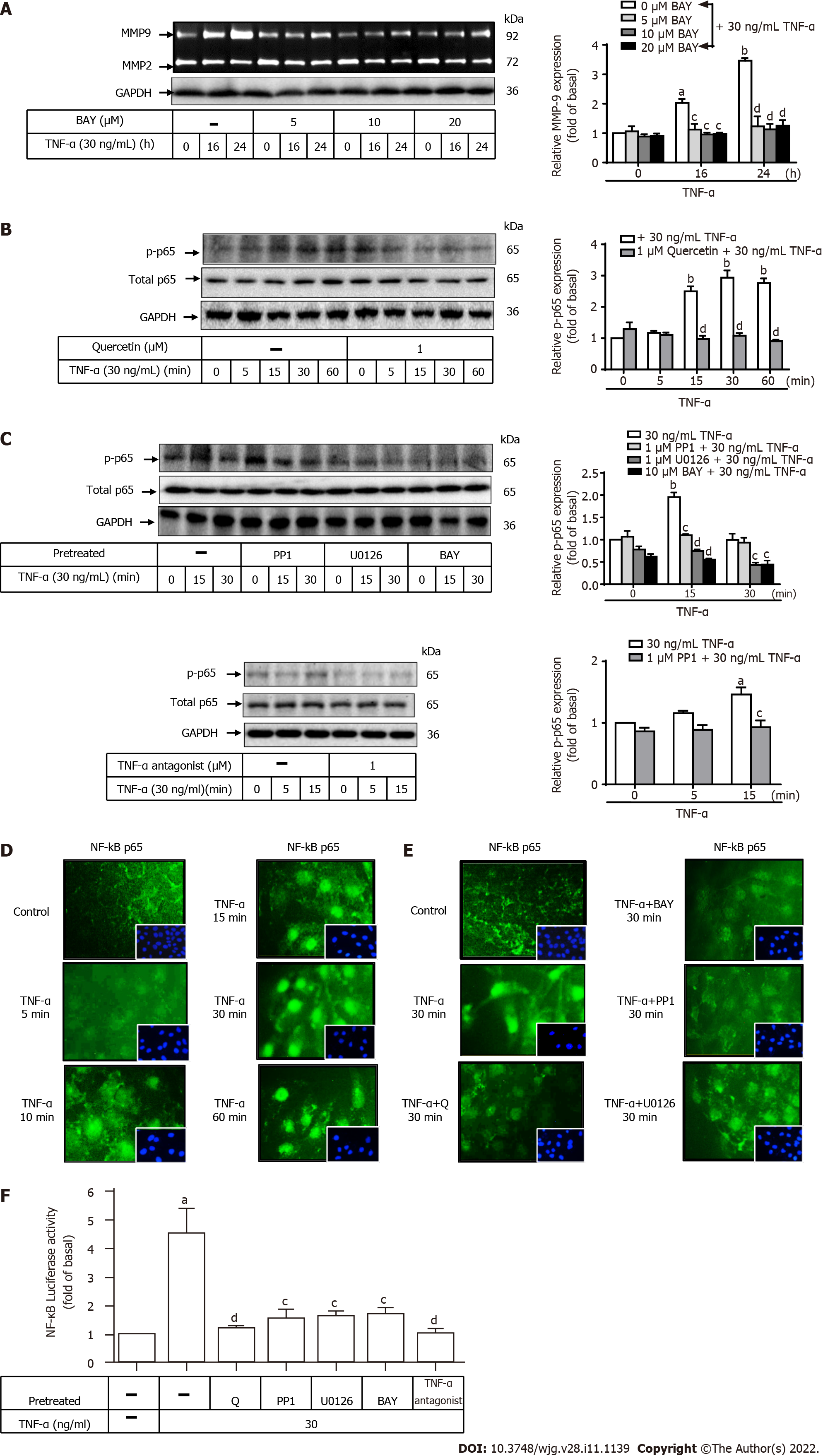
Figure 7 Quercetin suppresses tumor necrosis factor-α-induced tumor necrosis factor-α antagonist-c-Src–extracellular-signal-regulated kinase 1/2–nuclear factor kappa B (p65) activation in normal human gastric mucosa epithelial cells.
A: Normal human gastric mucosa epithelial cells (GES-1) were either untreated or treated with BAY (0, 5, 10, or 20 mM) for 0, 16, or 24 h; compounds were added 1 h before the addition of tumor necrosis factor-α (TNF-α) (30 ng/mL) as described in the Materials and Methods section. Matrix metallopeptidase-9 zymogen activity was measured by gelatin zymography. B and C: Cells were either untreated or treated with quercetin (B; 0 or 1 mM), PP1 (C; 0 or 1 mM), U0126 (0 or 1 mM), BAY (0 or 10 mM), or TNF-α antagonist (1 mM) for 1 h before the addition of TNF-α (30 ng/mL) and incubated for 0, 16, or 24 h. Nuclear factor kappa B (NF-κB) phosphorylation was examined by Western blot analysis; D and E: GES-1 cells were treated with 30 ng/mL TNF-α for 0, 5, 10, 15, 30, or 60 min (D) or quercetin (1 mM), PP1 (1 mM), U0126 (1 mM), or BAY (10 mM) for 1 h (E) before exposure to TNF-α (30 ng/mL) for 30 min; F: GES-1 cells were transfected with the human NF-κB–Luc response element reporter plasmids containing the dual-luciferase reporter system. The cells were then pretreated with PP1 (0 or 1 mM), U0126 (0 or 1 mM), BAY (0 or 10 mM), quercetin (1 mM), or TNF-α antagonist (1 mM) for 1 h and exposed to TNF-α for 1 h. Then, luciferase activity was measured. Firefly luciferase activity was standardized to that of Renilla luciferase. One-way ANOVA was used for comparisons among different treatment time points (aP < 0.05, bP < 0.01 vs control cells in 0 h). One-way ANOVA was used for comparisons among different treatments simultaneously (cP < 0.05, dP < 0.01 vs TNF-α-stimulated cells). Data are expressed as the mean ± SEM of three independent experiments. ERK: Extracellular-signal-regulated kinase; GAPDH: Glyceraldehyde-3-phosphate dehydrogenase; GES-1: Normal human gastric mucosa epithelial cell line; MMP-9: Matrix metallopeptidase-9; NF-κB: Nuclear factor kappa B; Q: Quercetin; TNF-α: Tumor necrosis factor-α.
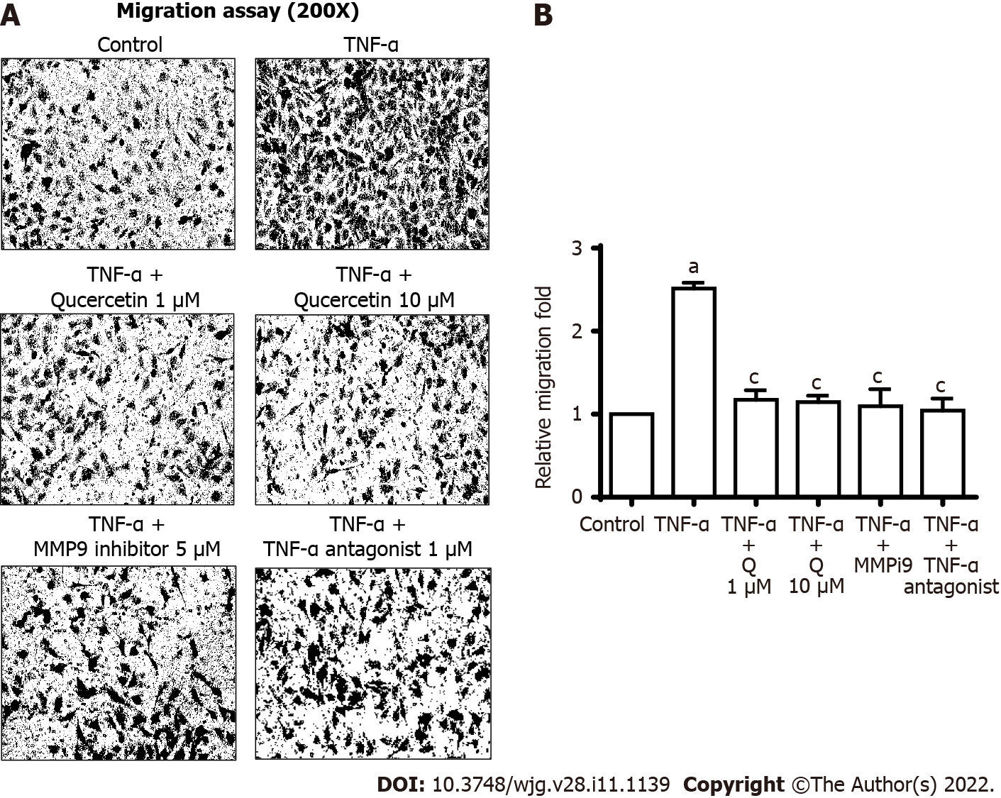
Figure 8 Quercetin exhibit antimetastatic activities in vitro.
A: Cells were plated in 24-well culture plates, grown to confluence, and starved with serum-free medium for 24 h. Cells were pretreated with a tumor necrosis factor-α (TNF-α) antagonist (1 mM), matrix metallopeptidase-9 inhibitor (5 mM), or quercetin (1 or 10 mM) for 1 h, and monolayer cells were incubated with TNF-α (30 ng/mL) for 24 h as described in the Materials and Methods section; B: One-way ANOVA was used for comparisons between the two groups (aP < 0.05, bP < 0.01 vs control cells). One-way ANOVA was used for comparisons among different treatments (cP < 0.05, dP < 0.01 vs TNF-α-stimulated cells). Data are expressed as the mean ± SEM of three independent experiments. GES-1: Normal human gastric mucosa epithelial cell line; MMP-9: Matrix metallopeptidase-9; MMP9i: MMP-9 inhibitor; TNF-α: Tumor necrosis factor-α.
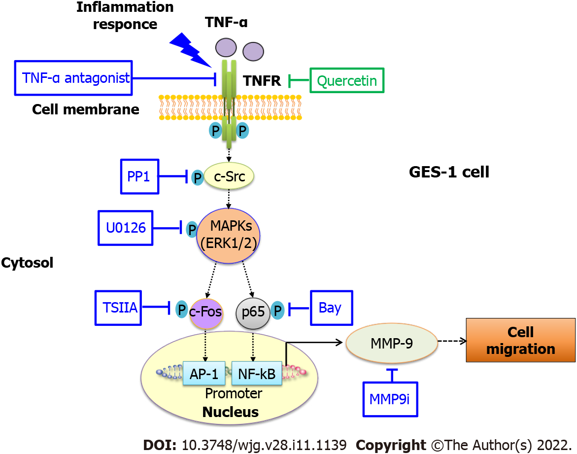
Figure 9 Schematic representation of the effects of quercetin on tumor necrosis factor-α-induced matrix metallopeptidase-9 expression and cell migration in normal human gastric mucosa epithelial cells.
Schematic diagram of the signaling pathways for quercetin-mediated attenuation of tumor necrosis factor-α (TNF-α)-induced inflammation via downregulation of matrix metallopeptidase-9 (MMP-9) expression in normal human gastric mucosa epithelial cells. Quercetin attenuates TNF-α-induced MMP-9 expression in normal human gastric mucosa epithelial cells through the proinflammatory TNFR-c-Src–extracellular-signal-regulated kinase 1/2–c-Fos and nuclear factor kappa B pathways. AP-1: Activator protein-1; c-Src: Proto-oncogene tyrosine-protein kinase Src; ERK: Extracellular-signal-regulated kinase; GES-1: Normal human gastric mucosa epithelial cell line; MAPK: Mitogen-activated protein kinase; MMP-9: Matrix metallopeptidase-9; MMP9i: MMP-9 inhibitor; NF-κB: Nuclear factor kappa B; TNF-α: Tumor necrosis factor-α; TSIIA: Tanshinone IIA.
- Citation: Hsieh HL, Yu MC, Cheng LC, Chu MY, Huang TH, Yeh TS, Tsai MM. Quercetin exerts anti-inflammatory effects via inhibiting tumor necrosis factor-α-induced matrix metalloproteinase-9 expression in normal human gastric epithelial cells. World J Gastroenterol 2022; 28(11): 1139-1158
- URL: https://www.wjgnet.com/1007-9327/full/v28/i11/1139.htm
- DOI: https://dx.doi.org/10.3748/wjg.v28.i11.1139


