Copyright
©The Author(s) 2021.
World J Gastroenterol. Nov 28, 2021; 27(44): 7649-7660
Published online Nov 28, 2021. doi: 10.3748/wjg.v27.i44.7649
Published online Nov 28, 2021. doi: 10.3748/wjg.v27.i44.7649
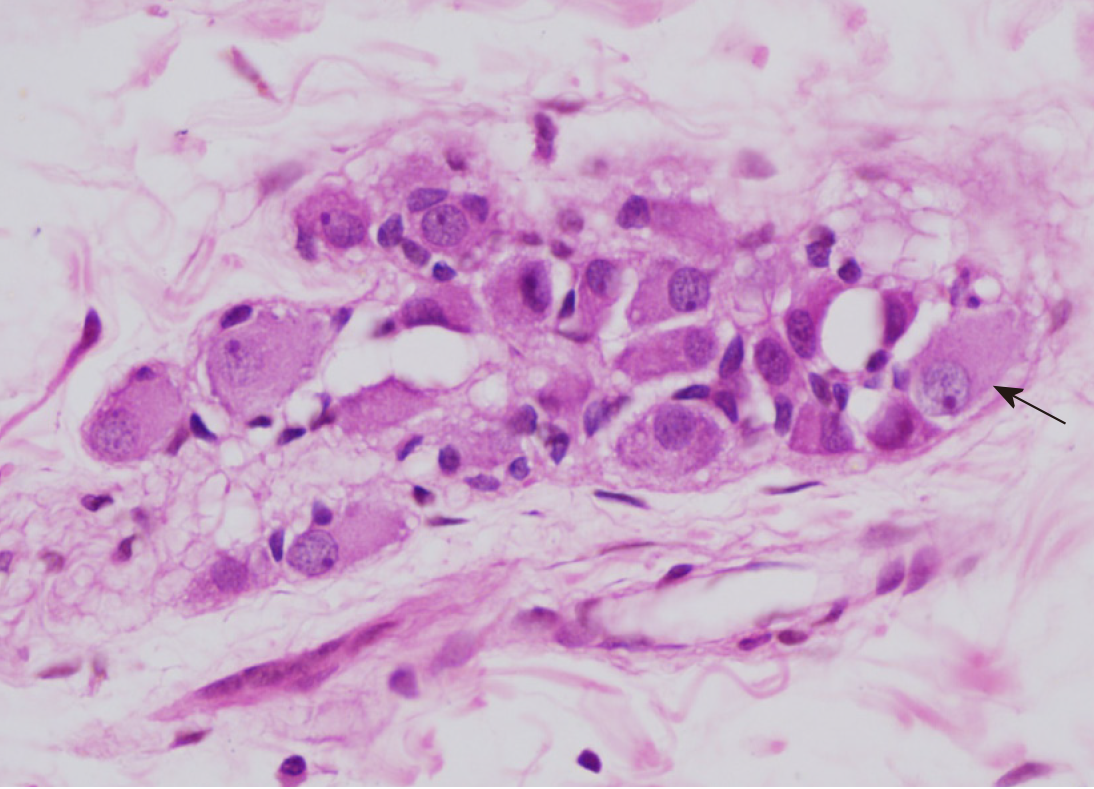
Figure 1 Intestinal neuronal dysplasia type B.
Submucosal nerve plexus with hyperganglionosis: giant ganglion. Ganglion cell (arrow) (H&E, 400 ×).
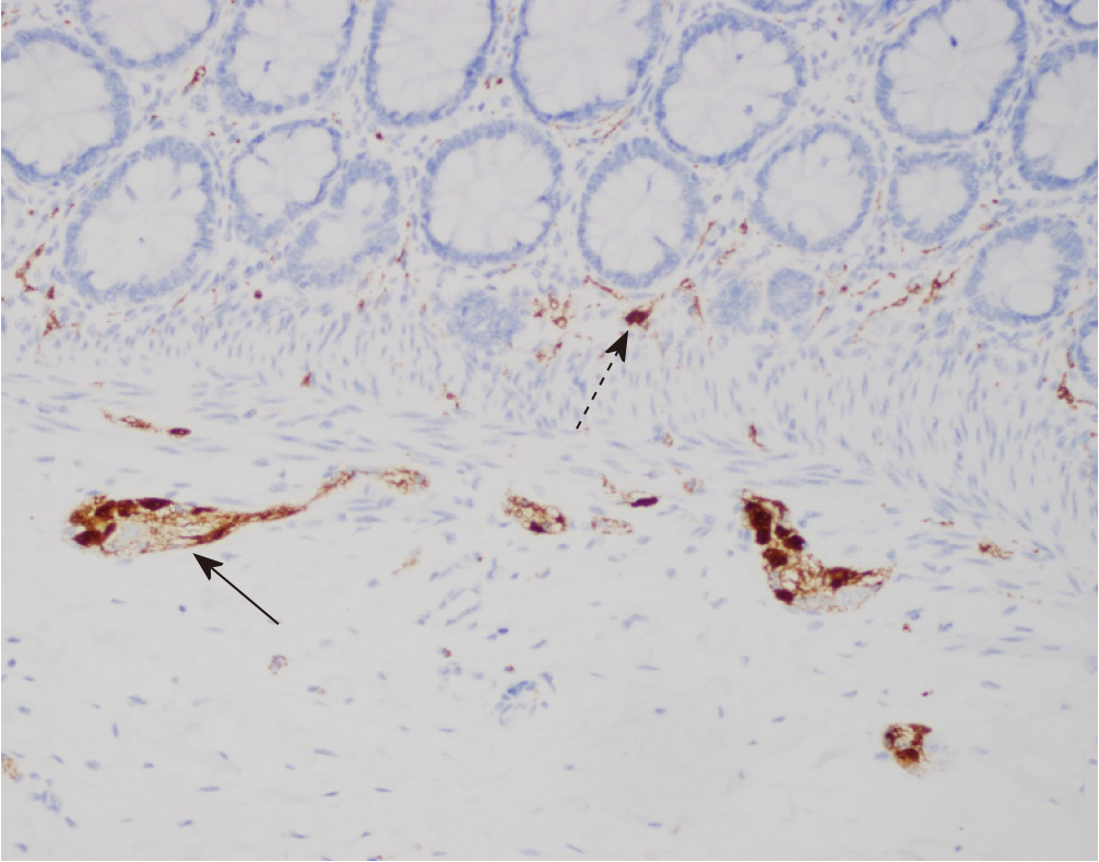
Figure 2 Calretinin immunohistochemistry in intestinal neuronal dysplasia type B.
Positive nuclear calretinin staining in neurons from a submucosal nerve plexus (arrow). Heterotopic neuron in the muscularis mucosa (dotted arrow) (200 ×).
- Citation: Terra SA, Gonçalves AC, Lourenção PLTA, Rodrigues MAM. Challenges in the diagnosis of intestinal neuronal dysplasia type B: A look beyond the number of ganglion cells. World J Gastroenterol 2021; 27(44): 7649-7660
- URL: https://www.wjgnet.com/1007-9327/full/v27/i44/7649.htm
- DOI: https://dx.doi.org/10.3748/wjg.v27.i44.7649


