Copyright
©The Author(s) 2021.
World J Gastroenterol. Nov 14, 2021; 27(42): 7376-7386
Published online Nov 14, 2021. doi: 10.3748/wjg.v27.i42.7376
Published online Nov 14, 2021. doi: 10.3748/wjg.v27.i42.7376
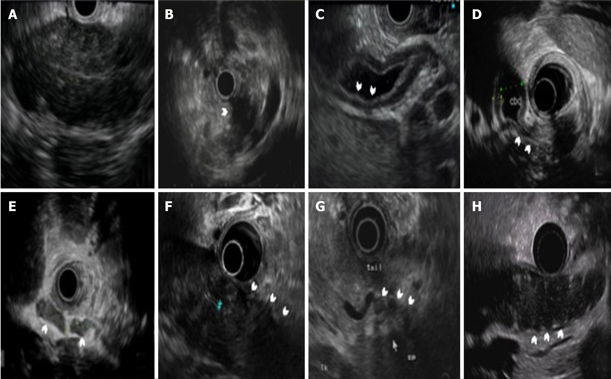
Figure 1 The typical endoscopic ultrasound findings in autoimmune pancreatitis.
The white arrows show the following: A: Diffuse hypoechoic area of the pancreatic body; B: Focal hypoechoic area of the pancreatic body; C: Thickened wall of the common bile duct; D: Intrapancreatic bile duct stenosis; E: Peripancreatic lymphadenopathy; F: Peripancreatic hypoechoic margin; G: Peripancreatic vessel involvement with stenosis of the splenic vein; H: Lobular outer margin.
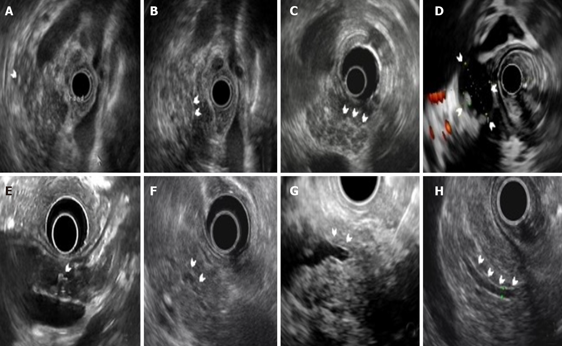
Figure 2 The pancreatic parenchymal and duct changes of chronic pancreatitis in autoimmune pancreatitis.
The white arrows show the following: A: Multiple hyperechoic foci; B: Hyperechoic strands in pancreatic head; C: Multiple lobularities with honeycombing; D: Cystic lesion connecting to the main pancreatic duct (MPD); E: MPD stone with acoustic shadow; F: Diffuse irregularity change of the MPD; G: Focal stenosis and upstream dilation of the MPD; H: Hyperechoic duct margin.
- Citation: Zhang SY, Feng YL, Zou L, Wu X, Guo T, Jiang QW, Wang Q, Lai YM, Tang SJ, Yang AM. Endoscopic ultrasound features of autoimmune pancreatitis: The typical findings and chronic pancreatitis changes. World J Gastroenterol 2021; 27(42): 7376-7386
- URL: https://www.wjgnet.com/1007-9327/full/v27/i42/7376.htm
- DOI: https://dx.doi.org/10.3748/wjg.v27.i42.7376


