Copyright
©The Author(s) 2021.
World J Gastroenterol. Nov 14, 2021; 27(42): 7299-7310
Published online Nov 14, 2021. doi: 10.3748/wjg.v27.i42.7299
Published online Nov 14, 2021. doi: 10.3748/wjg.v27.i42.7299
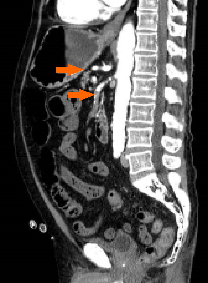
Figure 1 Abdominal computed tomography with intravenous contrast, sagittal scan showing thrombosis of the superior mesenteric artery and the common hepatic artery (arrows).
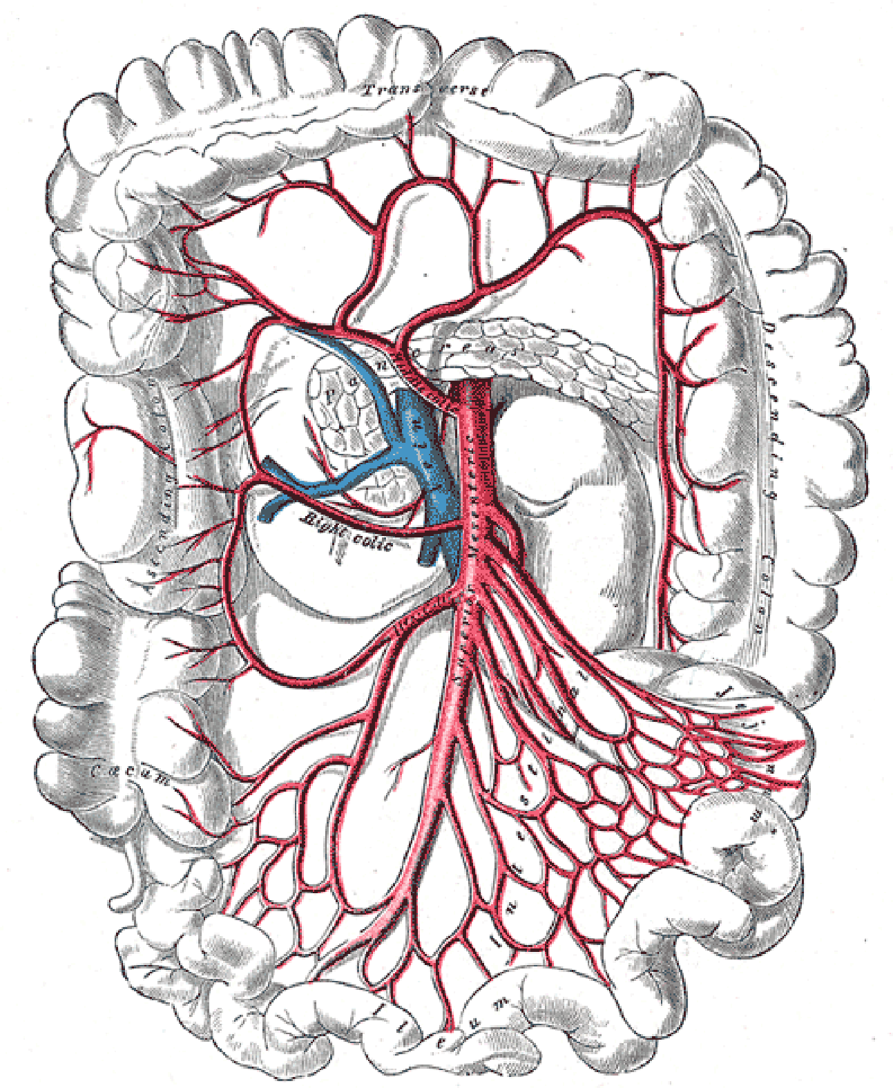
Figure 2 Splanchnic vascular anatomy, detail of colonic arteries (Case courtesy of Assoc Prof Craig Hacking, Radiopaedia.
org, rID: 54523.
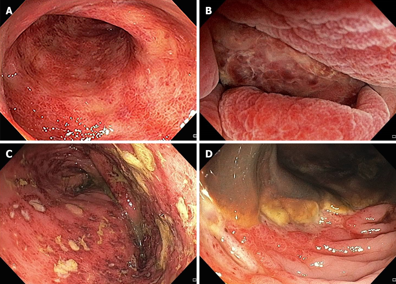
Figure 3 Endoscopic signs of ischemia, showing moderate diffuse erythema (A), severe erythema with mucosal edema and erosions (B), multiple ulcerations and inflammatory exudate (C), necrosis (D).
- Citation: Sadalla S, Lisotti A, Fuccio L, Fusaroli P. Colonoscopy-related colonic ischemia. World J Gastroenterol 2021; 27(42): 7299-7310
- URL: https://www.wjgnet.com/1007-9327/full/v27/i42/7299.htm
- DOI: https://dx.doi.org/10.3748/wjg.v27.i42.7299


