Copyright
©The Author(s) 2021.
World J Gastroenterol. Nov 14, 2021; 27(42): 7240-7246
Published online Nov 14, 2021. doi: 10.3748/wjg.v27.i42.7240
Published online Nov 14, 2021. doi: 10.3748/wjg.v27.i42.7240
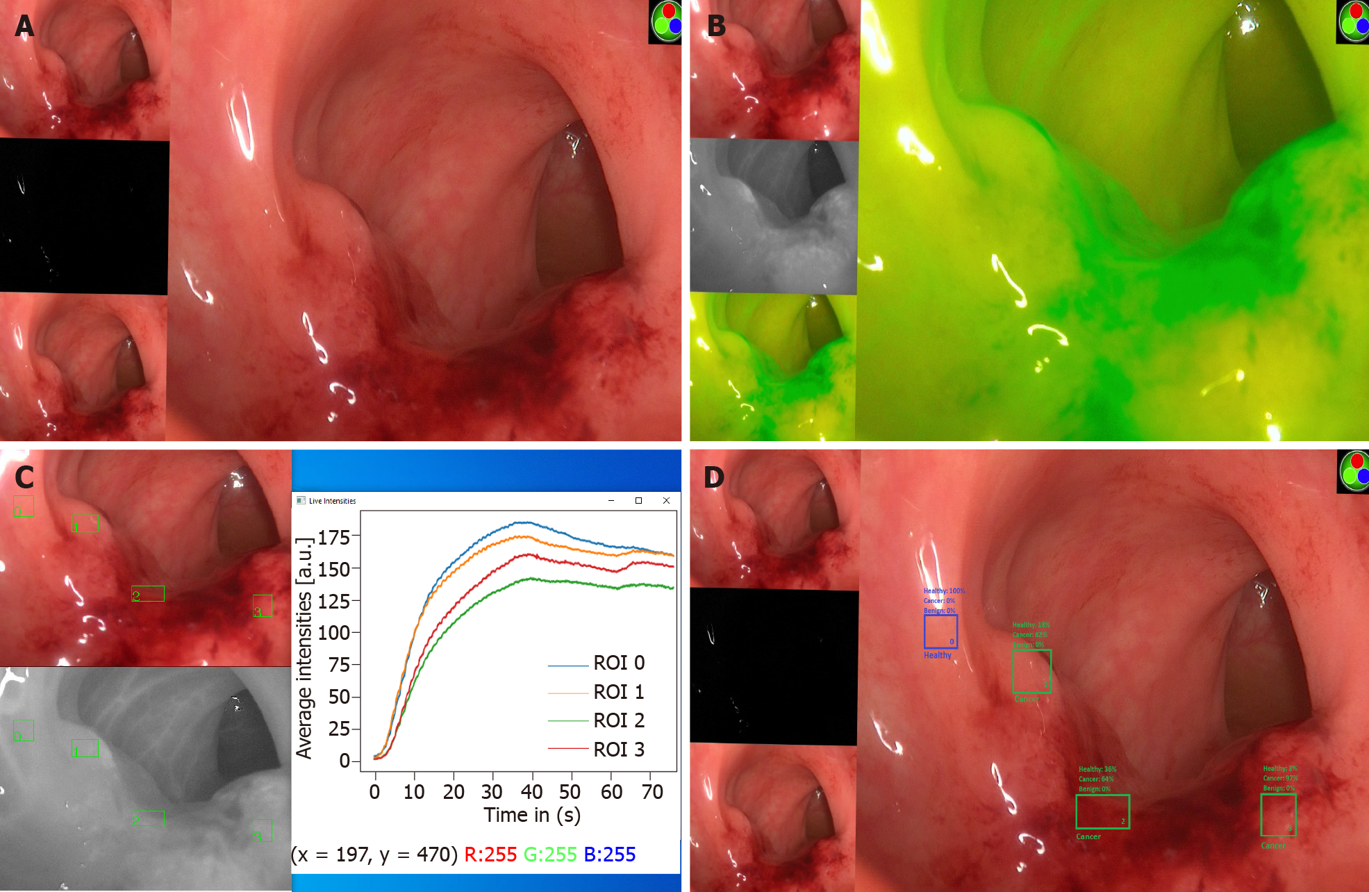
Figure 1 Digital surgery in action–real-time discrimination of tissue nature using fluorescence imaging and artificial intelligence.
A: Transanal imaging of a rectal lesion using a Pinpoint (Novadaq, Mississauga, Canada; Stryker, Kalamazoo, MI, United States) near infrared imaging system; B: Following intravenous administration of indocyanine green (ICG) (0.25 mg/kg), fluorescence is observed within the lesion and surrounding tissue; C: In tandem with the ICG administration, regions of interest within healthy and unhealthy tissue (regions 0–3) are chosen by the surgeon for real-time assessment. Image tracking is performed using the visible light mode and light intensity readings extracted from the corresponding regions within the infrared spectrum video for each of these regions over time; D: Intensity profiles are subsequently fitted to biophysics-inspired artificial intelligence models of fluid movement in tissue to predict tissue nature with a binary outcome (healthy vs cancer) and a probability score (%).
- Citation: Hardy NP, Cahill RA. Digital surgery for gastroenterological diseases. World J Gastroenterol 2021; 27(42): 7240-7246
- URL: https://www.wjgnet.com/1007-9327/full/v27/i42/7240.htm
- DOI: https://dx.doi.org/10.3748/wjg.v27.i42.7240


