Copyright
©The Author(s) 2021.
World J Gastroenterol. Aug 14, 2021; 27(30): 5060-5075
Published online Aug 14, 2021. doi: 10.3748/wjg.v27.i30.5060
Published online Aug 14, 2021. doi: 10.3748/wjg.v27.i30.5060
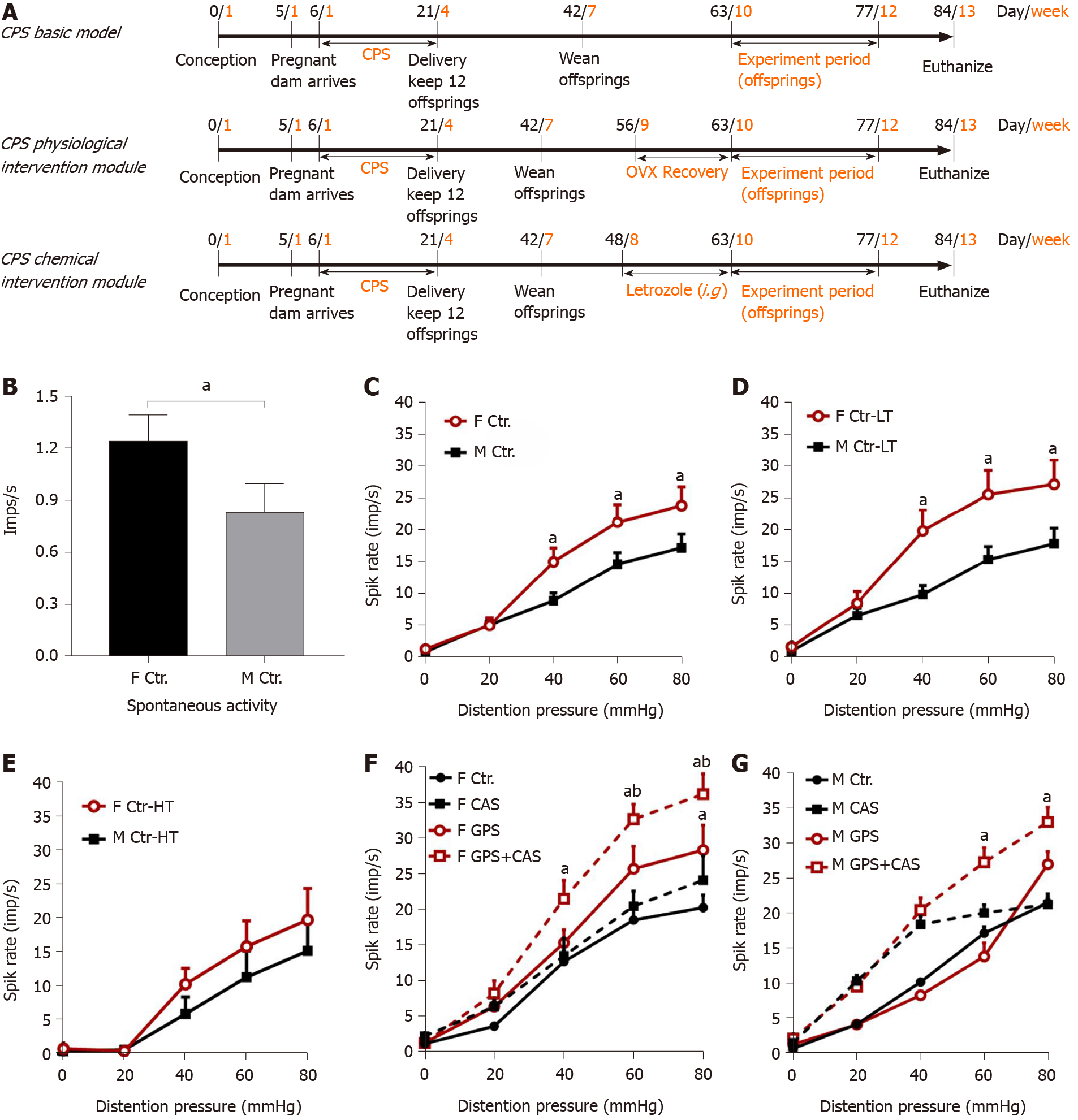
Figure 1 Primary afferent responses to colorectal distention.
A: Chronic prenatal stress (CPS) plus chronic adult stress (CAS) model. Pregnant dams were subjected to prenatal stress from on day 11 of gestation. Ovariectomy (OVX) or sham surgery was performed on female prenatal-stress offspring on day 56. Daily Letrozole was initiated on day 49, 2 wk prior to initiation of adult stress. Treatment was continued through the stress protocol; B: Spontaneous activity (SA) of single afferent units in male and female control rats (n = 70 fibers in 6 rats in each group, t-test, aP < 0.05); C: Average response to graded colorectal distention (CRD) of 56 afferent fibers in 6 male and 70 afferent fibers in 6 female control rats; two-way analysis of variance (ANOVA; aP < 0.05 vs the same pressure male group); D: Responses of low-threshold (LT) fibers to CRD in 42 fibers in 6 male rats and 40 fibers in 6 female control rats (ANOVA, aP < 0.05 vs the same pressure male group); E: Responses of high-threshold (HT) afferent fibers to CRD in 14 fibers in 6 male and 29 fibers in 6 female control rats; F: Effects of CAS on afferent fiber responses to CRD from 59 fibers in 6 control and 99 fibers in 6 CPS female rats; (two-way ANOVA, aP < 0.05 vs the same pressure control group, bP < 0.05 vs the same pressure CPS group); G: Effects of CAS on afferent fiber responses to CRD in control and CPS male rats (n = 6 rats, 57 fibers for control and 95 fibers for CPS female group; two-way ANOVA, aP < 0.05 vs the same pressure-control group).
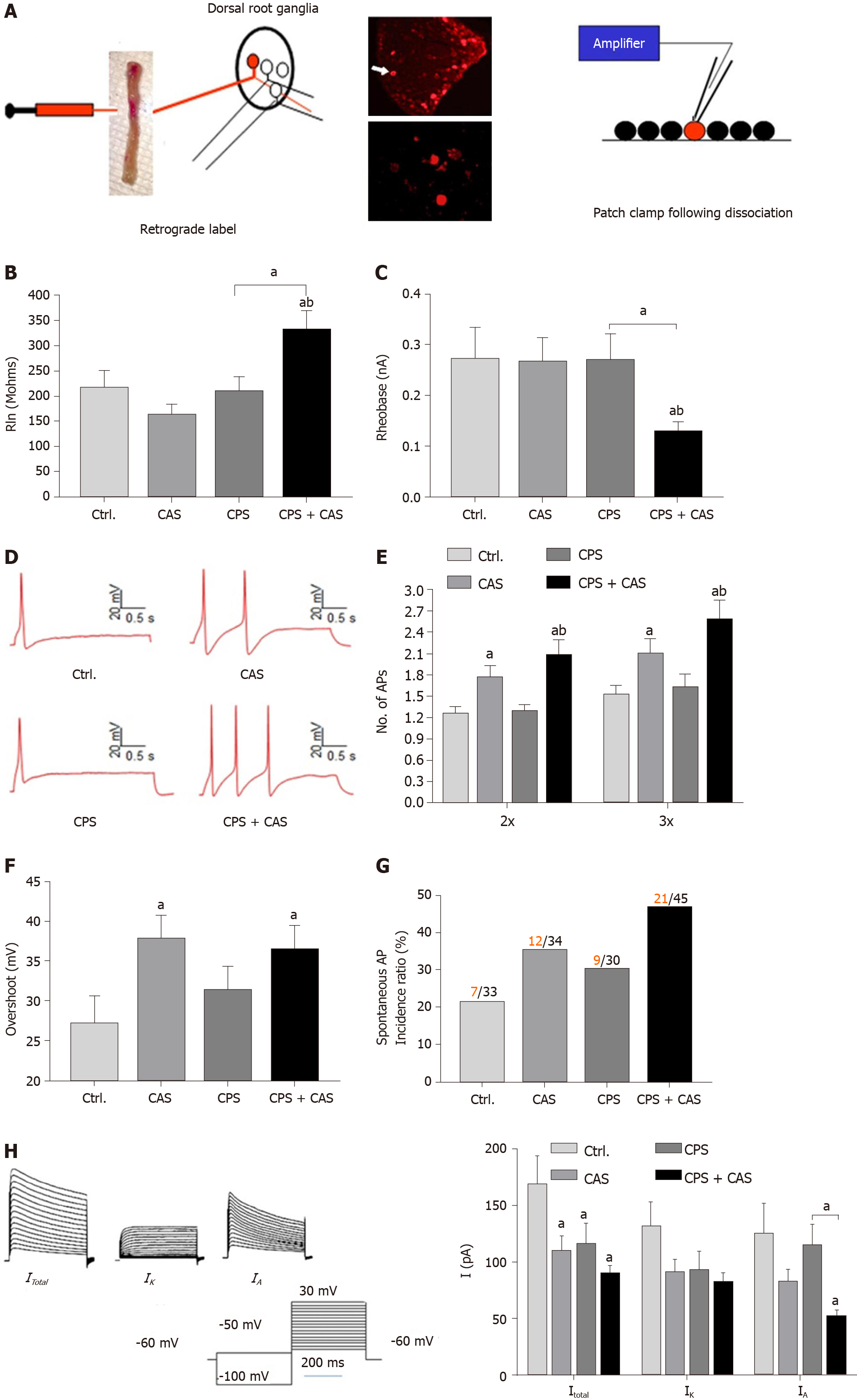
Figure 2 Patch clamp recording in colonic dorsal root ganglion neurons from female rats.
A: Patch clamp process of cell labeling. Under isoflurane anesthesia, the lipid soluble fluorescent dye 9-DiI was injected into muscularis externa of the exposed distal colon (left figure). Lumbosacral (L6–S2) dorsal root ganglions (upper photograph) were isolated and DiI-labeled neurons were identified by fluorescence microscopy (lower photograph). Electrophysiological properties of each neuron were measured using whole-cell current and voltage clamp protocols (right figure); B: Rheobase from all four experimental groups (n = 5 rats, 45 cells in each group, one-way ANOVA, aP < 0.05 vs control or bP < 0.05 vs CAS); C: Representative action potentials (APs) elicited by current injection at 2 × the rheobase in neurons from control, chronic adult stress (CAS), chronic prenatal stress (CPS) and CPS + CAS female rats; D: Membrane input resistance from all four groups (n = 5 rats, 45 cells in each group, one-way ANOVA, aP < 0.05 vs control; bP < 0.05 vs CPS); E: Number of APs elicited by current injection at either 2 × and 3 × the rheobase in all four experimental groups (two-way ANOVA, aP < 0.05 vs control; bP < 0.05 vs CPS); F: AP overshoot recorded from all four experimental groups (aP < 0.05 vs control); G: The proportion of neurons from each experimental group exhibiting spontaneous APs. Red numbers represent spontaneous AP firing cells; black numbers represent total cells; H: Representative total, IK and IA current tracings and average values of potassium currents: Itotal, IK and IA are shown in female CPS + CAS, CAS, CPS (n = 15 neurons, from 5 rats in each group), and control groups (n = 12 neurons from 5 rats); two-way ANOVA, aP < 0.05 vs each control group.
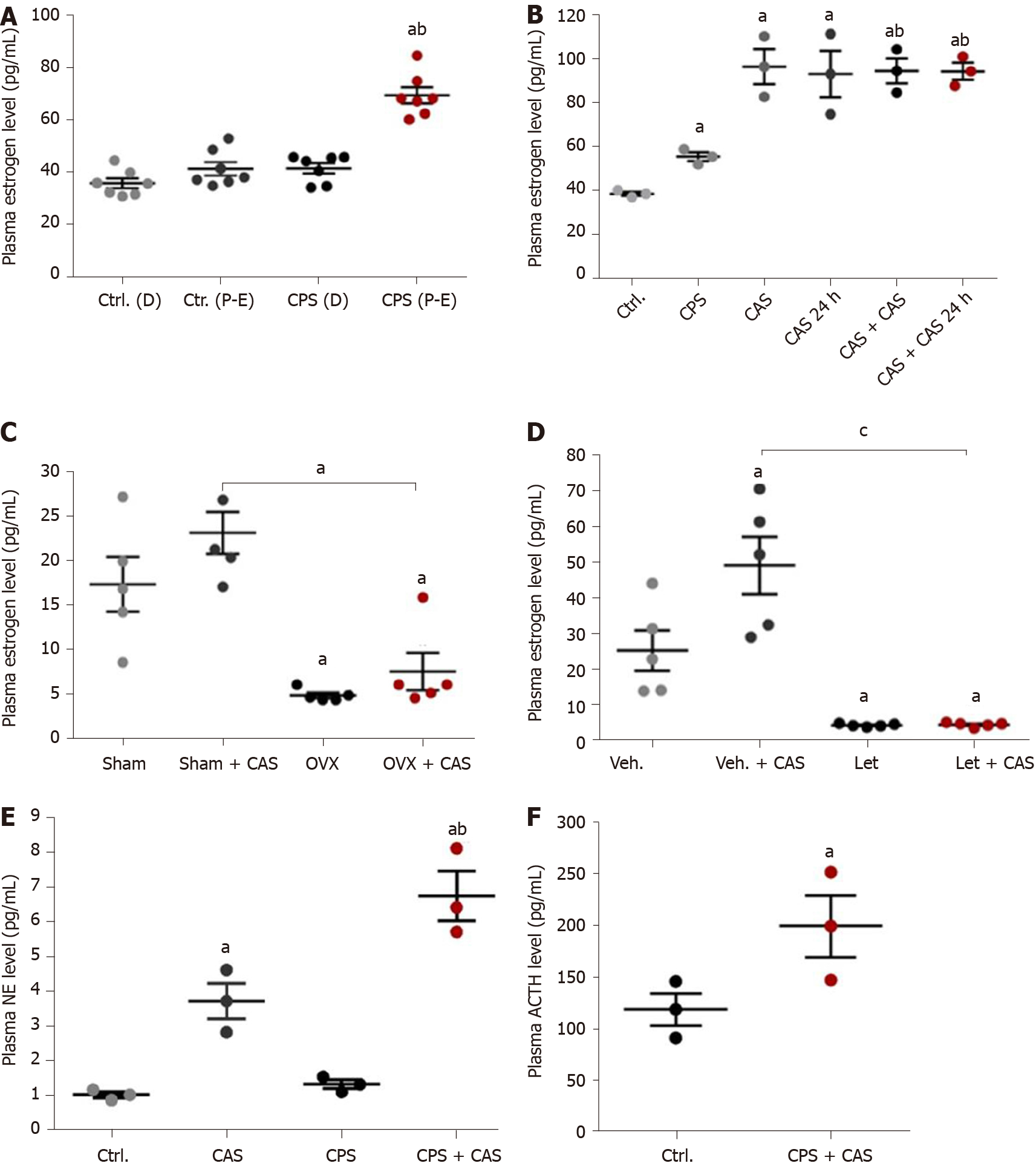
Figure 3 Effects of chronic prenatal stress, chronic adult stress, ovariectomy, and letrozole treatment on plasma estrogen levels in female rats.
A: Plasma estrogen level in control and chronic prenatal stress (CPS) rats by estrus cycle phase (n = 8 rats, one-way ANOVA, aP < 0.05 vs control proestrus/estrus (P-E) phase; bP < 0.05 vs CPS diestrus (D) phase); B: Plasma estrogen levels increased in CPS rats and following chronic adult stress (CAS) 24 h after the last adult stressor (n = 8 rats, one-way ANOVA, aP < 0.05 vs control; bP < 0.05 vs CPS); C: Ovariectomy (OVX) significantly reduced CPS female rat plasma estrogen levels before and after CAS (n = 5 rats, one-way ANOVA, aP < 0.05 vs sham group); D: Letrozole treatment significantly reduced CPS female rat plasma estrogen levels before or after CAS (n = 5 rats, one-way ANOVA, aP < 0.05 vs vehicle group; cP < 0.0001); E: Plasma norepinephrine levels from control, CAS, CPS and CPS + CAS group female rats (n = 5 rats, one-way ANOVA, aP < 0.05 vs control; bP < 0.05 vs CPS); F: Plasma adrenocorticotropic hormone (ACTH) levels from control and CPS + CAS group female rats (n = 5 rats, t-test, aP < 0.05 vs control).
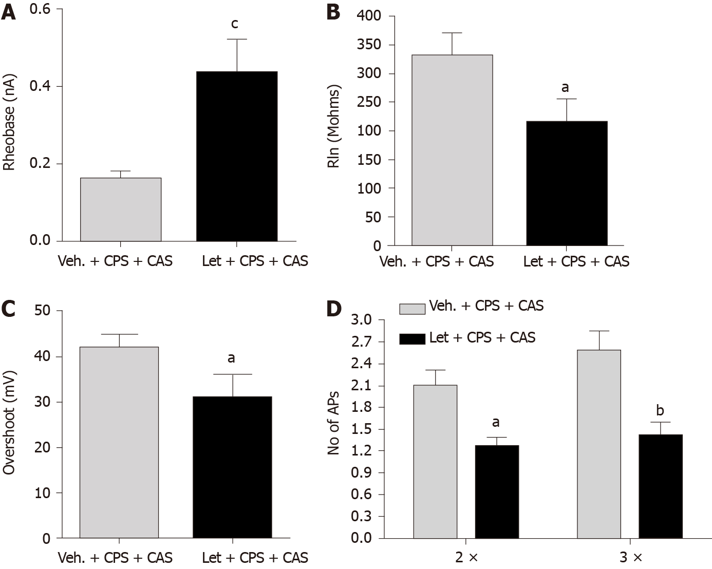
Figure 4 Effects of Letrozole treatment on colon dorsal root ganglion neuron excitability.
A: Rheobase (n = 45 cells in 6 rats in each group, t-test, cP < 0.001 vs Veh. + chronic adult stress [CAS] + chronic prenatal stress [CPS]); B: Membrane input resistance (RIn) (t-test, aP < 0.05); C: Action potential (AP) overshoot (t-test, aP < 0.05); D: Number of APs elicited by current injection at 2 × and 3 × rheobase (two-way ANOVA, aP < 0.05; bP < 0.01).
Figure 5 Brain-derived neurotrophic factor expression in lumbar-sacral spinal cord is regulated by estrogen.
A: Plasma estrogen levels in cycling females that received a bolus estradiol (E2) infusion on day 1; B: Lumbar-sacral spinal cord brain-derived neurotrophic factor (BDNF) mRNA following bolus estrogen infusion; C: Lumbar-sacral spinal cord BDNF protein following bolus estrogen infusion. (n = 8 rats in each group, two-way ANOVA, aP < 0.05 vs vehicle group).
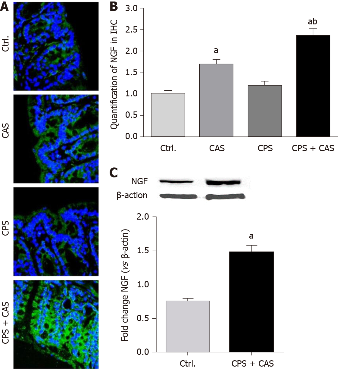
Figure 6 Nerve growth factor expression level in the colon wall.
A: Immunohistochemical staining of nerve growth factor (NGF; green) was detected with nuclear counterstaining staining (blue) in controls, chronic adult stress (CAS), chronic prenatal stress (CPS) and CPS + CAS group female rat colon walls. × 400 magnification representative pictures were shown; B: Quantification of NGF levels from colon wall in immunohistochemistry (IHC) (n = 4 rats in each group, one-way ANOVA, aP < 0.05 vs control group; bP < 0.05 vs CPS group); C: Western blots of NGF protein from control and CPS + CAS female rats colon wall tissue (n = 6 rats in each group, t-test, aP < 0.05 vs control group).
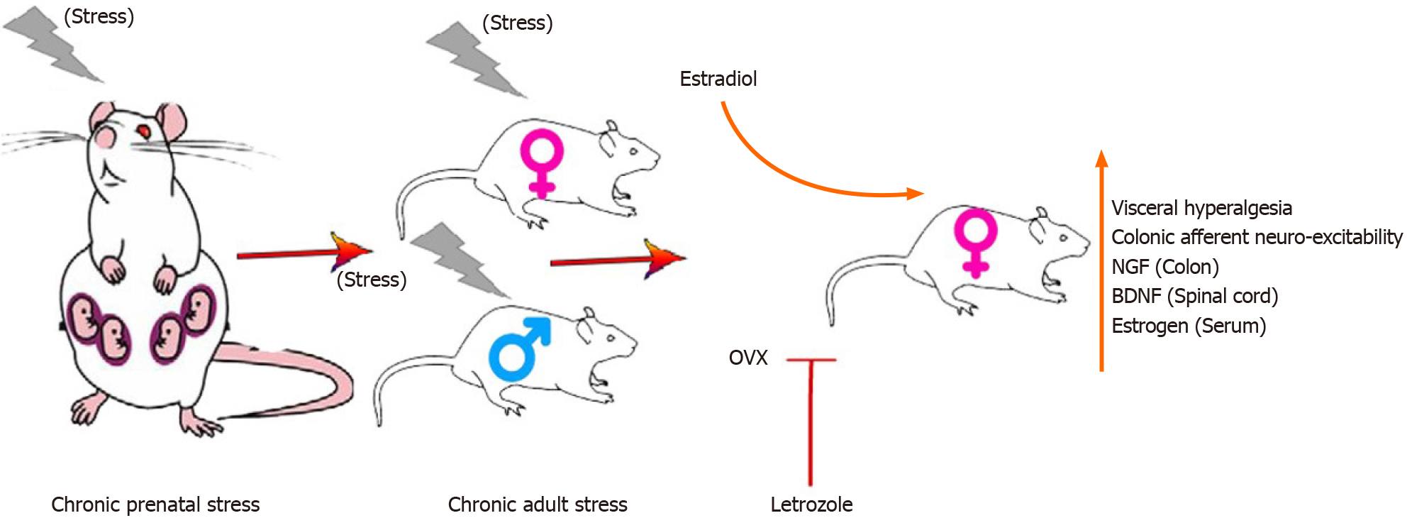
Figure 7 Summary diagram of estrogen re-enhanced visceral hyperalgesia investigated in chronic prenatal stress plus chronic adult stress models.
BDNF: Brain-derived neurotrophic factor; NGF: Nerve growth factor.
- Citation: Chen JH, Sun Y, Ju PJ, Wei JB, Li QJ, Winston JH. Estrogen augmented visceral pain and colonic neuron modulation in a double-hit model of prenatal and adult stress. World J Gastroenterol 2021; 27(30): 5060-5075
- URL: https://www.wjgnet.com/1007-9327/full/v27/i30/5060.htm
- DOI: https://dx.doi.org/10.3748/wjg.v27.i30.5060


