Copyright
©The Author(s) 2021.
World J Gastroenterol. Aug 14, 2021; 27(30): 5047-5059
Published online Aug 14, 2021. doi: 10.3748/wjg.v27.i30.5047
Published online Aug 14, 2021. doi: 10.3748/wjg.v27.i30.5047
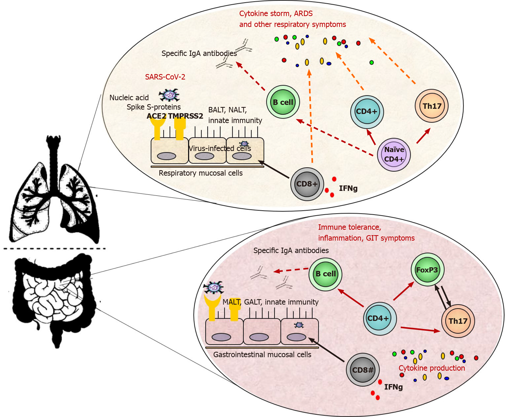
Figure 1 Nasal-, bronchial- and mucosa-associated lymphoid tissue are the first line of defense.
Airborne infections usually penetrate the upper airway mucosa, where a higher viral load is found. Nasopharynx-associated lymphoid tissue is involved in the induction of the immune response against the microorganisms by promoting the differentiation and activation of immune cells such as Th1- and Th2 cells, dendritic cells, macrophages, resident microfold M cells, innate lymphoid cells, immunoglobulin (Ig)A-switched B cells, as well as immune mediators and molecules (i.e. beta-defensins, galectins, collectins, cytokines). Similar immune processes are also observed in the gut mucosa. However, the expansion of CD4+ T-helper cells, CD8+ cytotoxic T cells, and plasma cells simultaneously with the ongoing innate immune response is critical for virus elimination. Additionally, specific secretory IgA (SIgA) plays an effective role in protection against severe acute respiratory syndrome coronavirus 2 by neutralization, inhibition of adherence, and agglutination. Additionally, SIgA does not activate the classical complement cascade pathway, and thus greater anti- than proinflammatory activity. Innate immune system and some innate receptors (PAMPS, DAMPS) are not shown for simplification of the figure. ACE2: Angiotensin-converting enzyme 2; ARDS: Acute respiratory distress syndrome; BALT: Bronchial-associated lymphoid tissue; GALT: Gut-associated lymphoid tissues; GIT: Gastrointestinal tract. IFN: Interferon; Ig: Immunoglobulin; MALT: Mucosa-associated lymphoid tissue; NALT: Nasopharynx-associated lymphoid tissue; SARS-CoV-2: Severe acute respiratory syndrome coronavirus 2; TMPRSS2: Transmembrane protease/serine subfamily member 2.
-
Citation: Velikova T, Snegarova V, Kukov A, Batselova H, Mihova A, Nakov R. Gastro
intestinal mucosal immunity and COVID-19. World J Gastroenterol 2021; 27(30): 5047-5059 - URL: https://www.wjgnet.com/1007-9327/full/v27/i30/5047.htm
- DOI: https://dx.doi.org/10.3748/wjg.v27.i30.5047


