Copyright
©The Author(s) 2021.
World J Gastroenterol. Jul 7, 2021; 27(25): 3705-3733
Published online Jul 7, 2021. doi: 10.3748/wjg.v27.i25.3705
Published online Jul 7, 2021. doi: 10.3748/wjg.v27.i25.3705
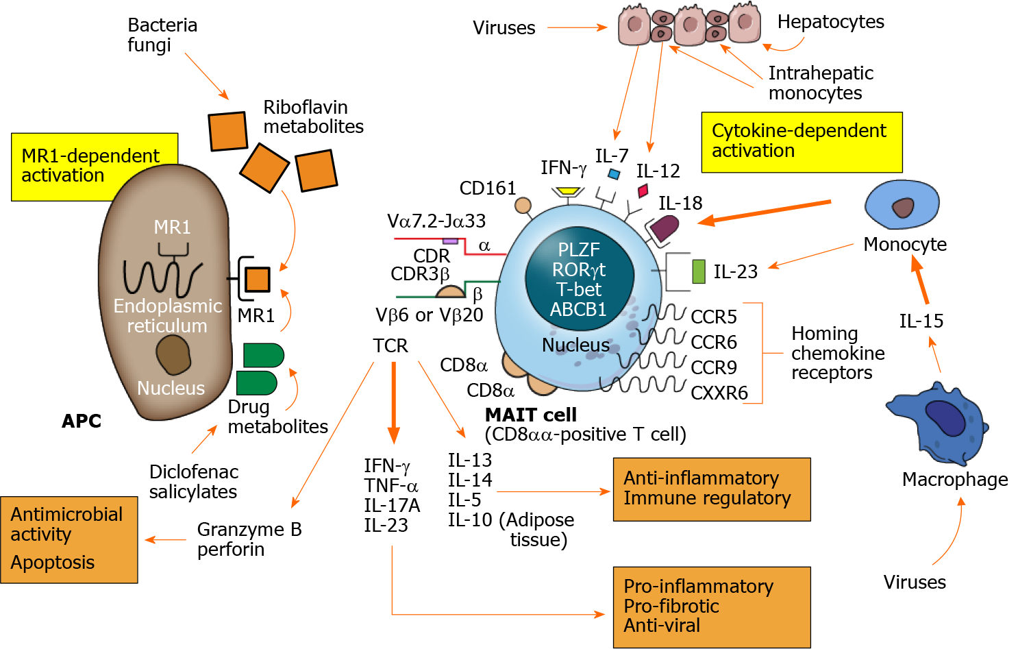
Figure 1 Mucosal-associated invariant T cell activation and actions.
Mucosal-associated invariant T (MAIT) cells are activated by MR1-dependent and cytokine-dependent mechanisms. The major histocompatibility complex class I-related protein, MR1, is expressed on the surface of the antigen presenting cell after stimulation. Riboflavin (vitamin B) metabolites synthesized by bacteria and fungi are presented by the MR1 molecule as are select drug metabolites. The T cell antigen receptor of the MAIT cell consists of a semi-invariant alpha (α) chain and restricted beta (β) chain with antigen selectivity influenced by short length complementarity-determining regions. MR1-dependent activation results in MAIT cell production of multiple cytokines as well as granzyme B and perforin. The cytokines can have pro-inflammatory, pro-fibrotic and antiviral effects (lower right panel) and anti-inflammatory and immune regulatory effects (lower right panel). The granzyme B and perforin can have antimicrobial activity and eliminate infected cells by apoptosis (lower left panel). Cytokine-dependent stimulation is activated by phagocytic macrophages and monocytes resident in the liver or circulation and by injured hepatocytes. Multiple cytokines can activate MAIT cells, especially interleukin 18, after virus infection. Chemokine receptors help direct the activated MAIT cells to the site of inflammation, and CXCR6 is the principal chemokine that directs MAIT cells to the liver. MAIT cells contain diverse nuclear transcription factors that influence phenotype and function, especially promyelocytic leukemia zinc finger, retinoic acid- related orphan receptor gamma t, and T-bet. The nucleus also contains the ABCB1 that affects resistance to gut-derived xenobiotics and certain drugs. The principal subset of activated MAIT cells consists of CD8αα-positive T cells. APC: Antigen presenting cell; TCR: T cell antigen receptor; CDR: Complementarity-determining region; IL: Interleukin; IFN-γ: Interferon-gamma; TNF-α: Tumor necrosis factor-alpha; MAIT: Mucosal-associated invariant T.
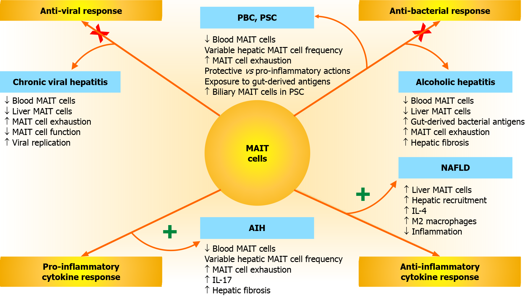
Figure 2 Mucosal-associated invariant T cell responses and associations with chronic liver disease.
Mucosal-associated invariant T (MAIT) cells have anti-viral, anti-bacterial, anti-inflammatory, and pro-inflammatory responses that can affect the occurrence, severity, and outcome of diverse chronic liver diseases, including chronic viral hepatitis, alcoholic hepatitis, non-alcoholic fatty liver disease (NAFLD), autoimmune hepatitis (AIH), primary biliary cholangitis (PBC), and primary sclerosing cholangitis (PSC). Impairments in the anti-viral and anti-bacterial responses of MAIT cells (indicated by an “X” across each activation pathway) may promote chronic viral hepatitis, PBC, PSC, and alcoholic hepatitis. Hyperactivity of the anti-inflammatory and pro-inflammatory cytokine responses of MAIT cells (indicated by a “+” next to each activation pathway) may affect NAFLD and AIH. Increased release of interleukin 4 (IL-4) and IL-17 may contribute to the modulation of these responses. MAIT: Mucosal-associated invariant T; NAFLD: Non-alcoholic fatty liver disease; AIH: Autoimmune hepatitis; PBC: Primary biliary cholangitis; PSC: Primary sclerosing cholangitis; IL: Interleukin.
- Citation: Czaja AJ. Incorporating mucosal-associated invariant T cells into the pathogenesis of chronic liver disease. World J Gastroenterol 2021; 27(25): 3705-3733
- URL: https://www.wjgnet.com/1007-9327/full/v27/i25/3705.htm
- DOI: https://dx.doi.org/10.3748/wjg.v27.i25.3705


