Copyright
©The Author(s) 2021.
World J Gastroenterol. Jun 21, 2021; 27(23): 3148-3157
Published online Jun 21, 2021. doi: 10.3748/wjg.v27.i23.3148
Published online Jun 21, 2021. doi: 10.3748/wjg.v27.i23.3148
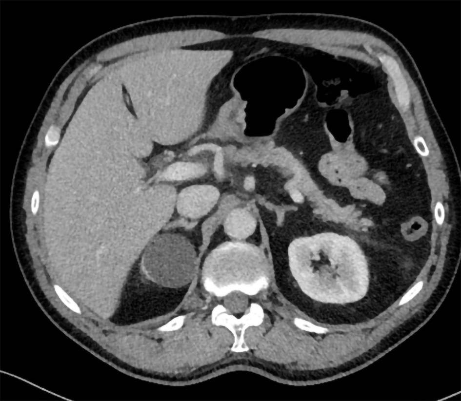
Figure 1 Abdominal computed tomography imaging of acute pancreatitis.
Inflammation is present around the head of the pancreas.
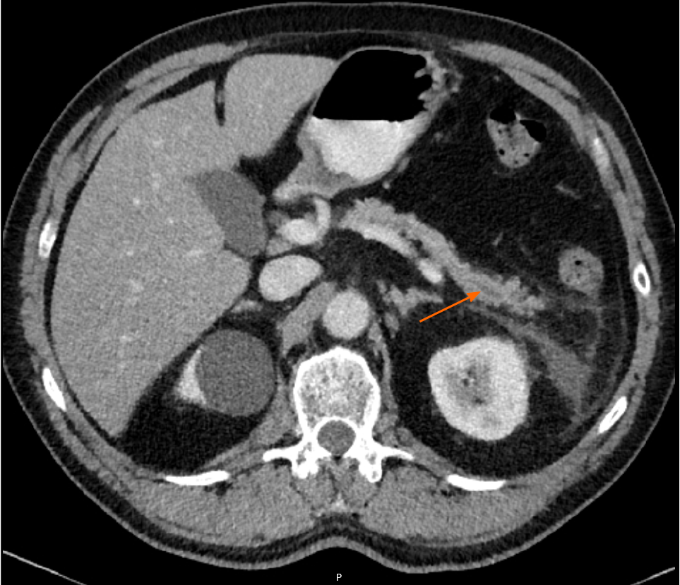
Figure 2 Abdominal computed tomography imaging of acute pancreatitis.
In the pancreatic tail, a dilatation of the pancreatic duct can be observed.
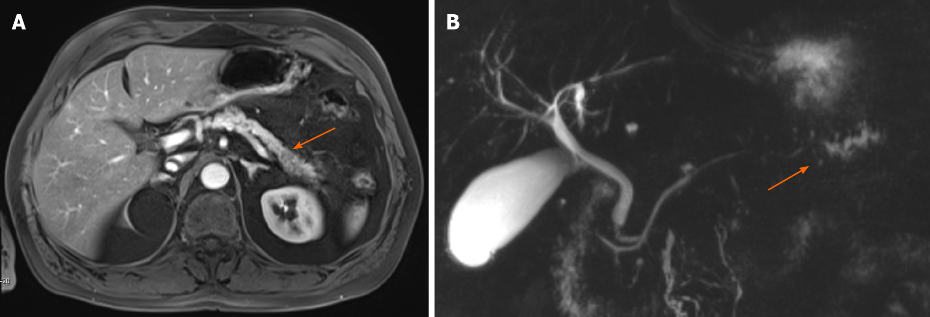
Figure 3 Abdominal magnetic resonance imaging of the pancreas and magnetic resonance cholangiopancreatography.
A: Abdominal magnetic resonance imaging of the pancreas; B: Magnetic resonance cholangiopancreatography. A hypo-intense lesion (A, arrow) is causing a pancreatic duct stenosis with upstream dilatation of the pancreatic duct (B, arrow).
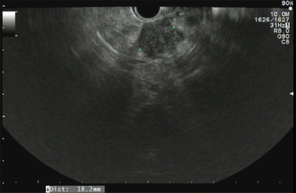
Figure 4 Endoscopic ultrasonography of the pancreas.
A hypo-echoic lesion measuring 18.2 mm is present. Biopsy of this lesion revealed a pancreatic ductal adenocarcinoma.
- Citation: Umans DS, Hoogenboom SA, Sissingh NJ, Lekkerkerker SJ, Verdonk RC, van Hooft JE. Pancreatitis and pancreatic cancer: A case of the chicken or the egg. World J Gastroenterol 2021; 27(23): 3148-3157
- URL: https://www.wjgnet.com/1007-9327/full/v27/i23/3148.htm
- DOI: https://dx.doi.org/10.3748/wjg.v27.i23.3148


