Copyright
©The Author(s) 2021.
World J Gastroenterol. May 14, 2021; 27(18): 2238-2250
Published online May 14, 2021. doi: 10.3748/wjg.v27.i18.2238
Published online May 14, 2021. doi: 10.3748/wjg.v27.i18.2238
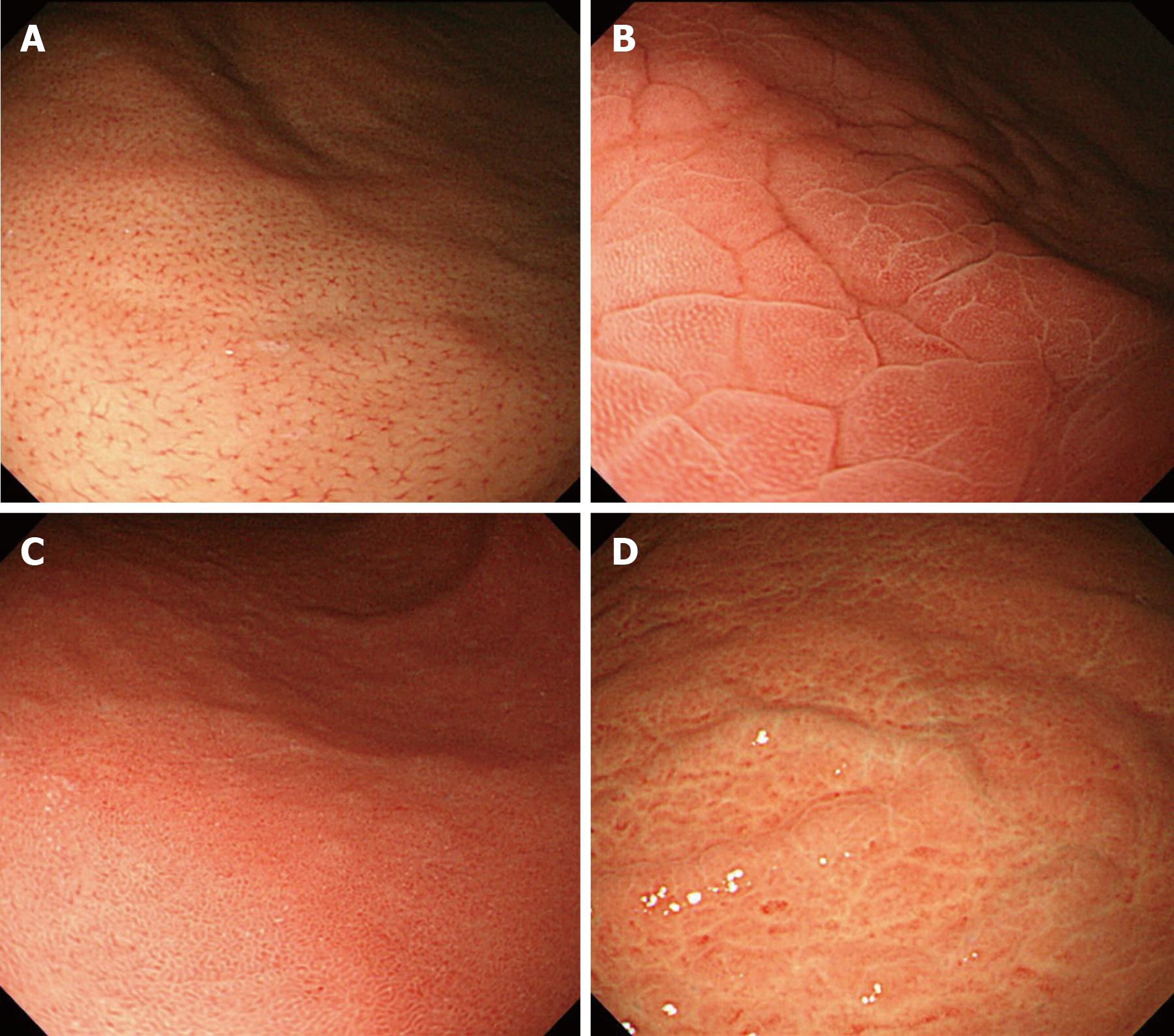
Figure 1 Normal and three abnormal mucosal patterns in the gastric corpus by standard endoscopy.
A: Normal pattern, numerous minute red dots; B: Type A, mosaic-like appearance; C: Type B, diffuse homogenous redness; D: Type C, irregular redness with grooves.
Figure 2 Micromucosal patterns in the gastric corpus observed by magnifying narrow-band imaging endoscopy.
A: Normal pattern characterized by regular arrangement of collecting venules and honeycomb-like subepithelial capillary network; B: Type Z-1, regular round pits with polygonal sulci; C: Type Z-2, more dilated and linear pits without sulci; D: Type Z-3, loss of gastric pits with coiled microvessels.
- Citation: Cho JH, Jeon SR, Jin SY, Park S. Standard vs magnifying narrow-band imaging endoscopy for diagnosis of Helicobacter pylori infection and gastric precancerous conditions. World J Gastroenterol 2021; 27(18): 2238-2250
- URL: https://www.wjgnet.com/1007-9327/full/v27/i18/2238.htm
- DOI: https://dx.doi.org/10.3748/wjg.v27.i18.2238


