Copyright
©The Author(s) 2021.
World J Gastroenterol. Apr 21, 2021; 27(15): 1655-1663
Published online Apr 21, 2021. doi: 10.3748/wjg.v27.i15.1655
Published online Apr 21, 2021. doi: 10.3748/wjg.v27.i15.1655
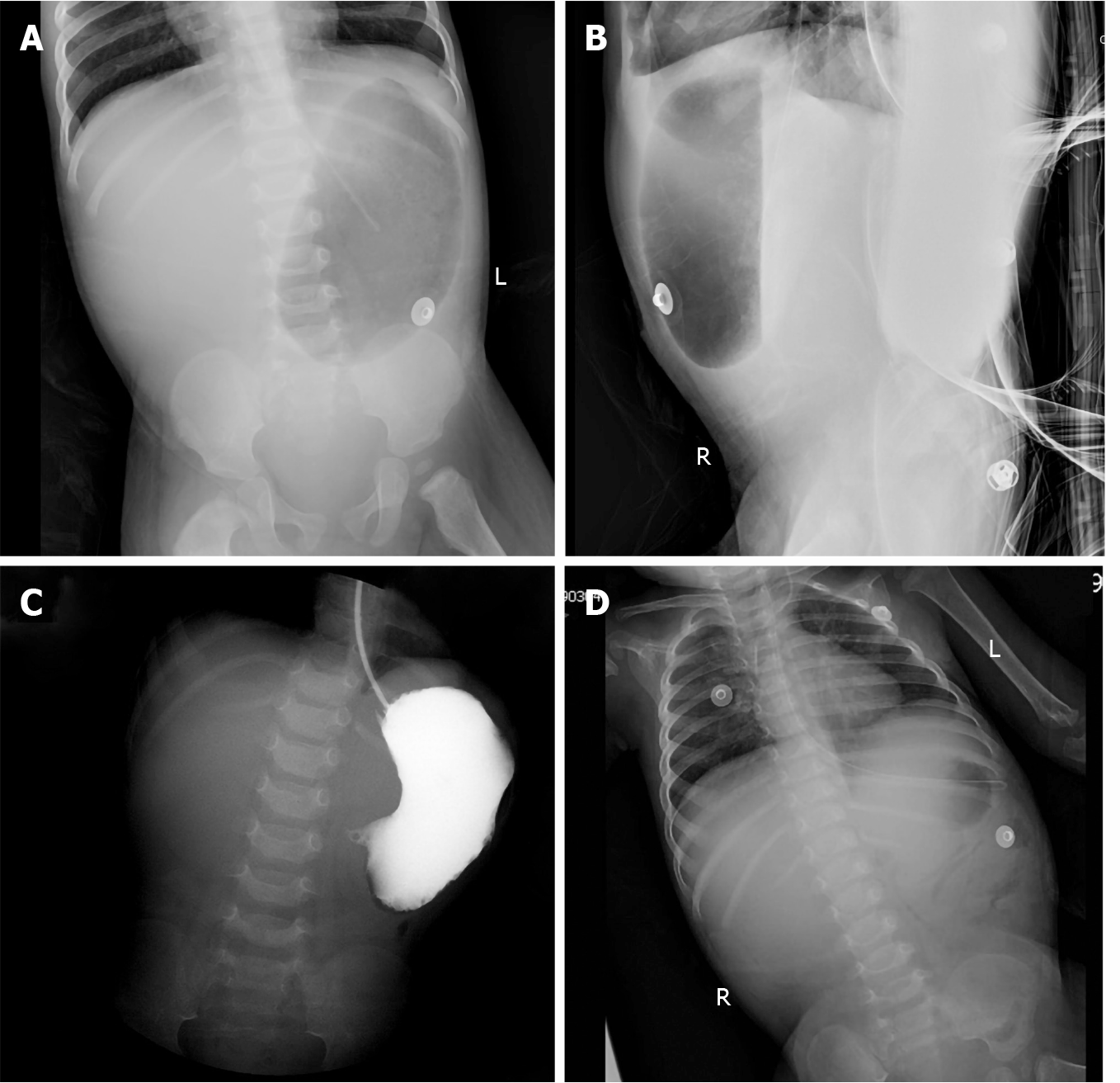
Figure 1 Abdominal plain.
A and B: Supine (A) and lateral (B) abdominal plain films showed a large stomach, and no free gas was found in the small intestine, colon, or rectum. A nasogastric tube was coiled in the stomach; C: The iopromide administered by gastric tube did not enter the duodenum; D: Re-examination of abdominal film revealed decreased gas in the stomach and a few inflatable intestines.
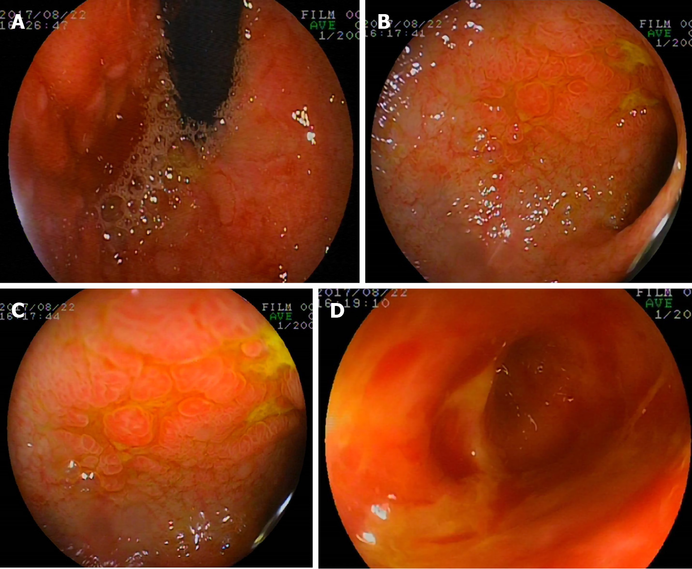
Figure 2 Gastroscopic findings.
A gastroscope was inserted into the horizontal part of the duodenum. Hyperemia, edema, lymphoid hyperplasia, focal ulceration, and pseudotumor formation were seen in the stomach and duodenum. A: Gastric fundus; B and C: Duodenal bulb; D: Dilatation of proximal duodenum.
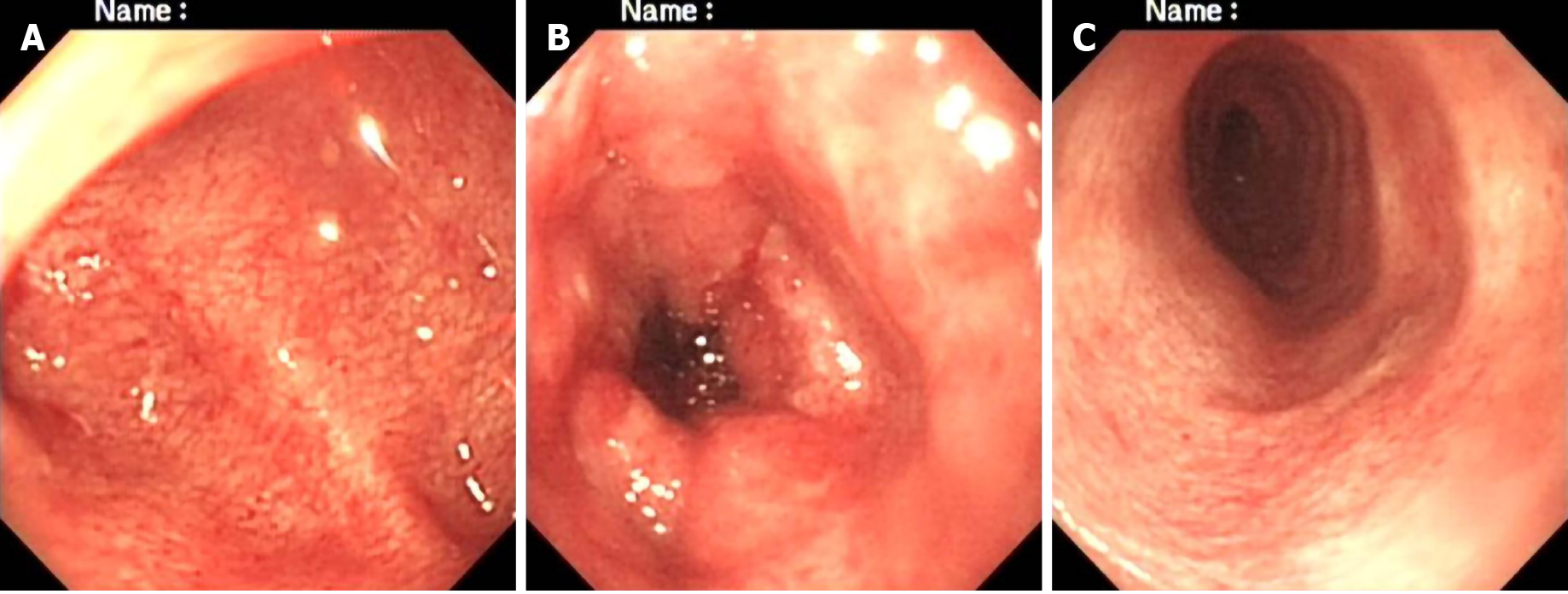
Figure 3 Colonoscopic findings.
A colonoscope reached the splenic flexure of the colon. It showed hyperemia, roughness, stiffness, friability, and easy bleeding of the mucosa. A: Rectum; B: Sigmoid colon; C: Descending colon.
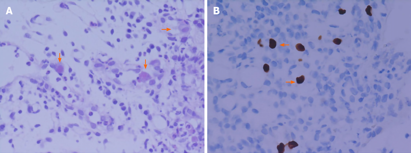
Figure 4 Gastrointestinal histopathologic findings.
Histopathologic photomicrographs of gastric, duodenal, descending colony-like, sigmoid colony-like, and rectal specimens. A: Inflammatory cell infiltration and characterized cytomegalic cells (arrows) containing eosinophilic intranuclear inclusions (hematoxylin and eosin: × 400); B: Owl’s eye inclusions (arrows) in cytomegalovirus-infected cells (immunohistochemical staining: × 400).
- Citation: Yang QH, Ma XP, Dai DL, Bai DM, Zou Y, Liu SX, Song JM. Gastrointestinal cytomegalovirus disease secondary to measles in an immunocompetent infant: A case report. World J Gastroenterol 2021; 27(15): 1655-1663
- URL: https://www.wjgnet.com/1007-9327/full/v27/i15/1655.htm
- DOI: https://dx.doi.org/10.3748/wjg.v27.i15.1655


