Copyright
©The Author(s) 2021.
World J Gastroenterol. Apr 21, 2021; 27(15): 1553-1562
Published online Apr 21, 2021. doi: 10.3748/wjg.v27.i15.1553
Published online Apr 21, 2021. doi: 10.3748/wjg.v27.i15.1553
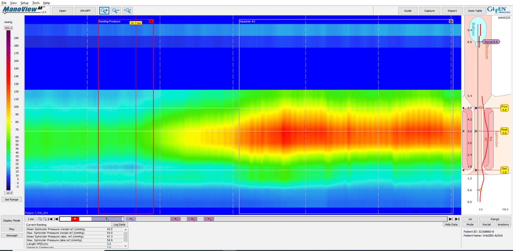
Figure 1 High-resolution anorectal manometry demonstrating normal resting and squeeze anal pressures.
Supplied from the Gastroenterology and Endoscopy United, The Nazareth Hospital, EMMS, Nazareth, Israel.
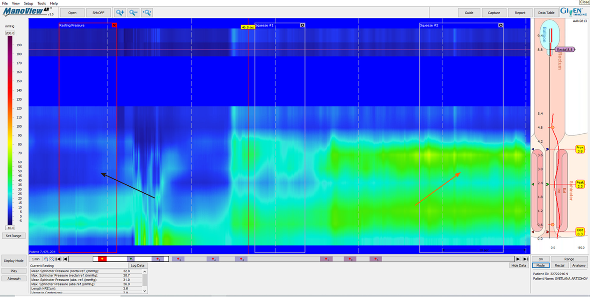
Figure 2 High-resolution anorectal manometry demonstrating low resting anal pressure (black arrow) and low squeeze pressure (orange arrow).
Supplied from the Gastroenterology and Endoscopy United, The Nazareth Hospital, EMMS, Nazareth, Israel.
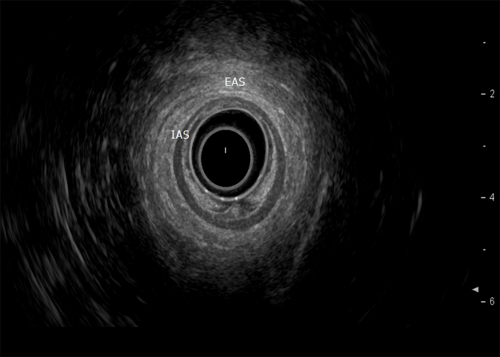
Figure 3 Normal internal and external anal sphincters (internal anal sphincter and external anal sphincter).
Supplied from the Department of Gastroenterology, Galilee Medical Center, Nahariya, Israel. EAS: External anal sphincter; IAS: Internal anal sphincter.
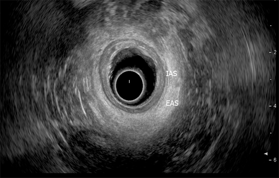
Figure 4 Internal anal sphincter atrophy manifested as sphincter thinning.
Supplied from the Department of Gastroenterology, Galilee Medical Center, Nahariya, Israel. EAS: External anal sphincter; IAS: Internal anal sphincter.
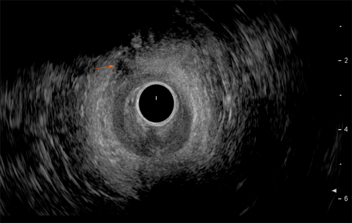
Figure 5 External anal sphincter tear.
Supplied from the Department of Gastroenterology, Galilee Medical Center, Nahariya, Israel.
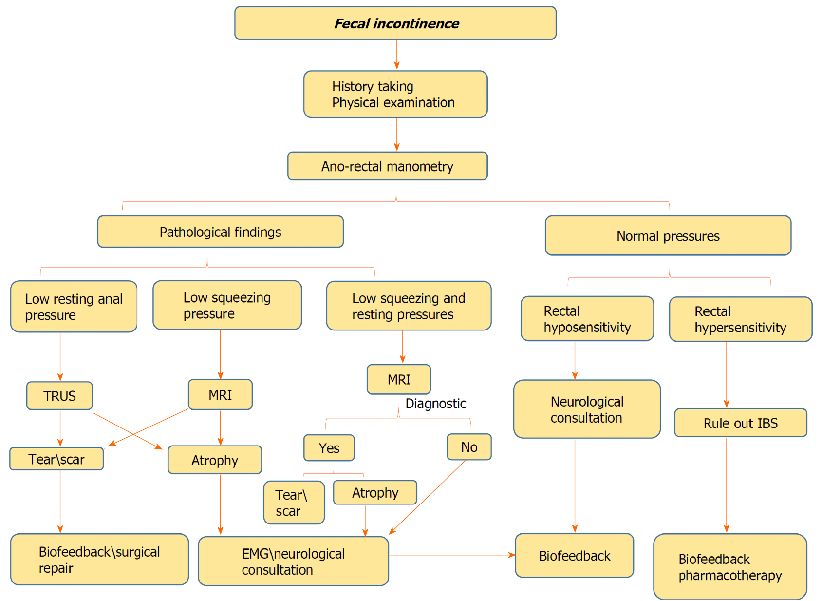
Figure 6 Stepwise approach of faecal incontinence investigation.
EMG: Electromyography; MRI: Magnetic resonance imaging; TRUS: Trans-rectal ultrasound.
- Citation: Sbeit W, Khoury T, Mari A. Diagnostic approach to faecal incontinence: What test and when to perform? World J Gastroenterol 2021; 27(15): 1553-1562
- URL: https://www.wjgnet.com/1007-9327/full/v27/i15/1553.htm
- DOI: https://dx.doi.org/10.3748/wjg.v27.i15.1553


