Copyright
©The Author(s) 2020.
World J Gastroenterol. Mar 7, 2020; 26(9): 973-983
Published online Mar 7, 2020. doi: 10.3748/wjg.v26.i9.973
Published online Mar 7, 2020. doi: 10.3748/wjg.v26.i9.973
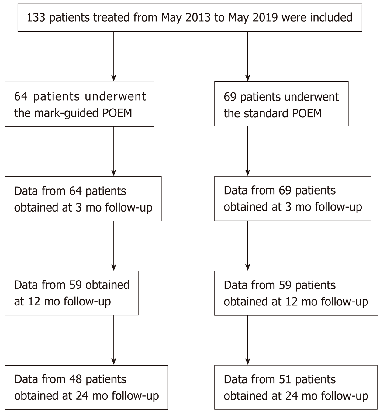
Figure 1 Flow chart.
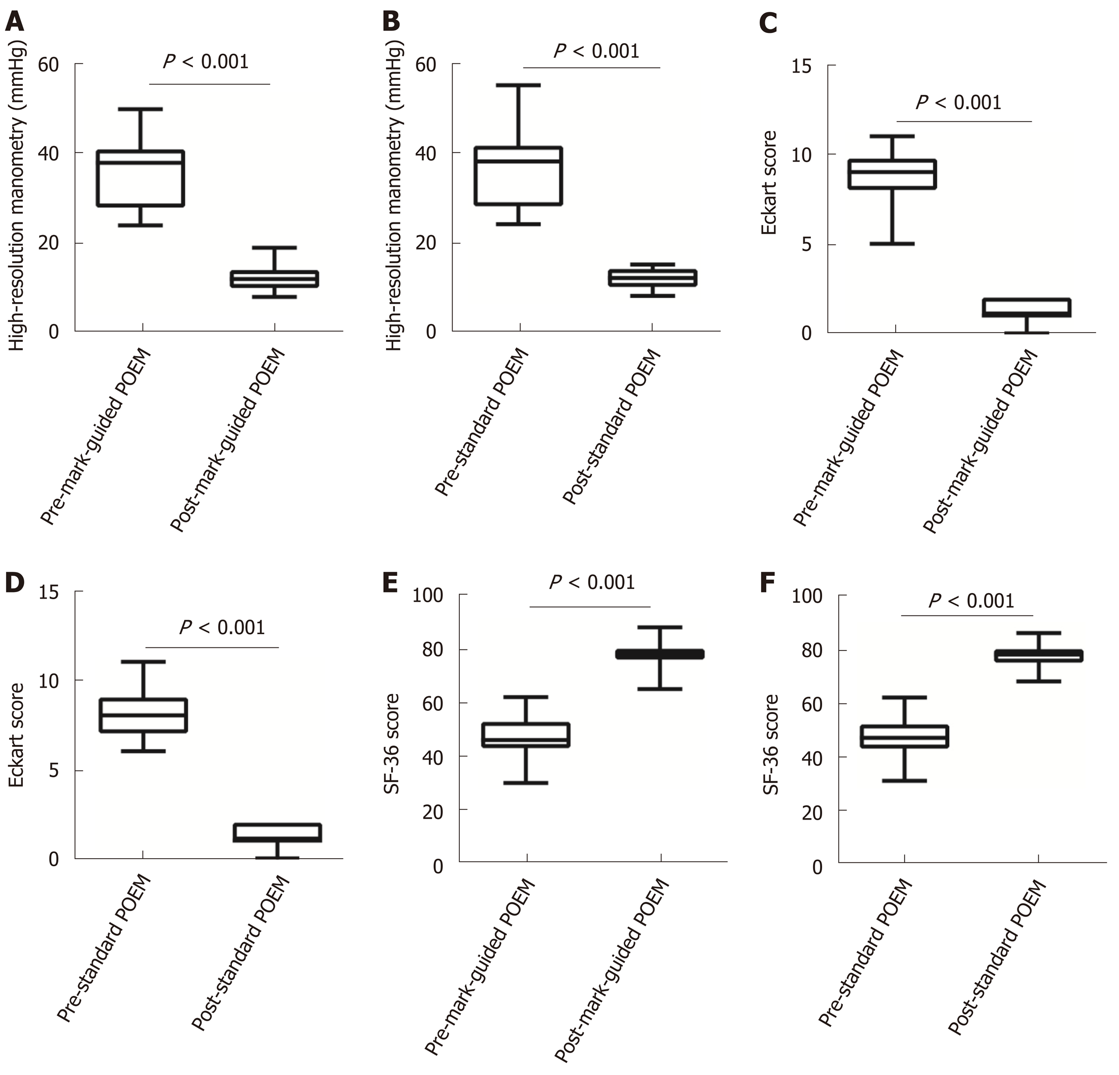
Figure 2 High-resolution manometry, Eckart score and 36-Item Short-Form Health Survey scores at 3-mo follow-up in the mark-guided peroral endoscopic myotomy group and standard peroral endoscopic myotomy group.
A-D: The pre-operative high-resolution manometry and Eckart scores were significantly decreased compared with the postoperative values in the two groups (all P < 0.001); E, F: The pre-operative 36-Item Short-Form Health Survey scores were significantly improved compared with the postoperative values in both groups (all P < 0.001).
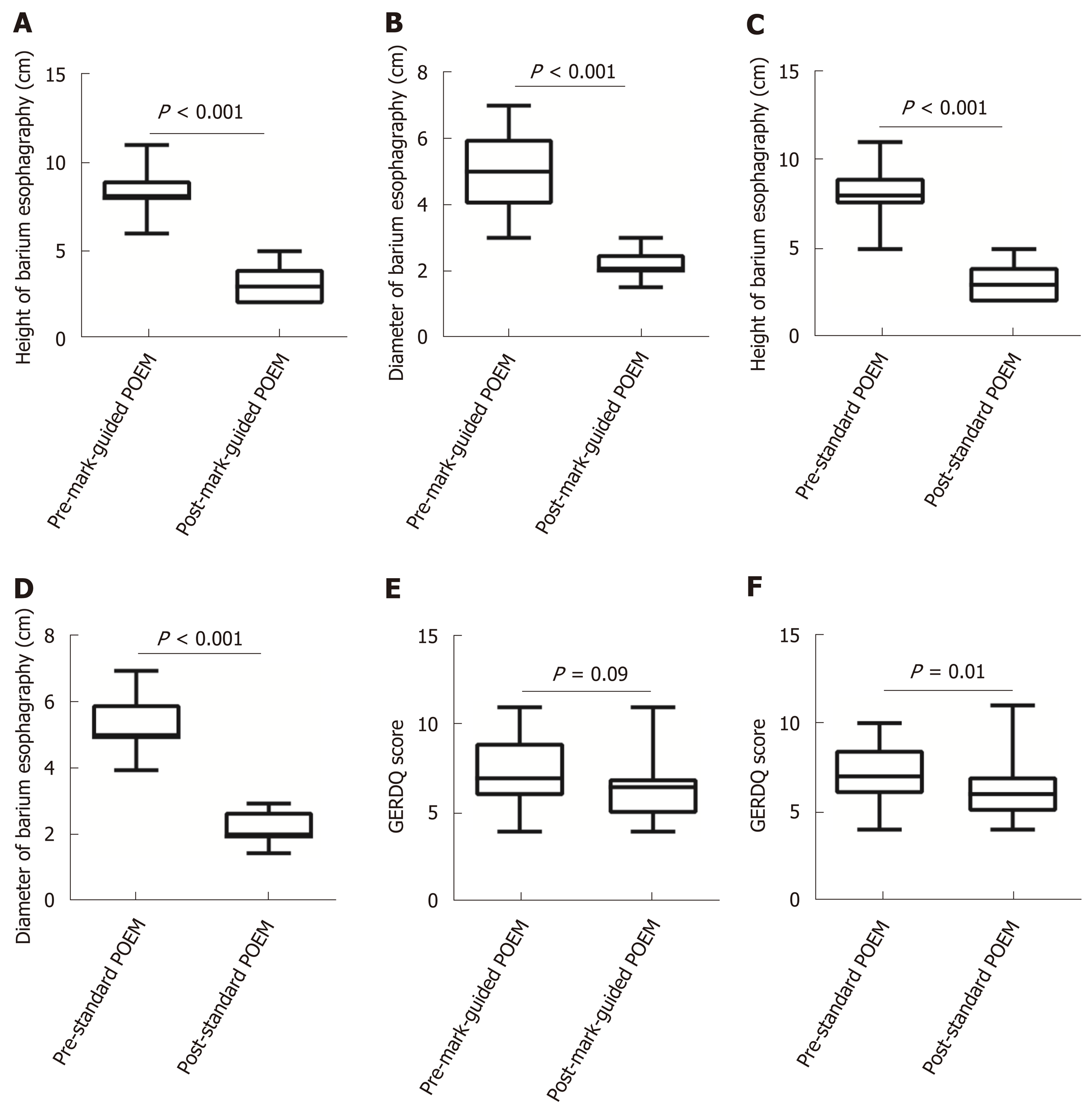
Figure 3 Barium esophagography at 3-mo follow-up in the mark-guided peroral endoscopic myotomy group and standard peroral endoscopic myotomy group.
A-D: The post-operative height and diameter of barium esophagography were significantly decreased compared with the pre-operative values in the two groups (all P < 0.001); E: The pre-operative Gastroesophageal reflux disease questionnaire score was significantly decreased compared with the post-operative value in the standard peroral endoscopic myotomy group (P = 0.01); F: No significant difference was observed between pre-operative and post-operative values in the mark-guided peroral endoscopic myotomy group (P = 0.09).
- Citation: Li DF, Xiong F, Yu ZC, Zhang HY, Liu TT, Tian YH, Shi RY, Lai MG, Song Y, Xu ZL, Zhang DG, Yao J, Wang LS. Effect and safety of mark-guided vs standard peroral endoscopic myotomy: A retrospective case control study. World J Gastroenterol 2020; 26(9): 973-983
- URL: https://www.wjgnet.com/1007-9327/full/v26/i9/973.htm
- DOI: https://dx.doi.org/10.3748/wjg.v26.i9.973


