Copyright
©The Author(s) 2020.
World J Gastroenterol. Feb 21, 2020; 26(7): 717-724
Published online Feb 21, 2020. doi: 10.3748/wjg.v26.i7.717
Published online Feb 21, 2020. doi: 10.3748/wjg.v26.i7.717
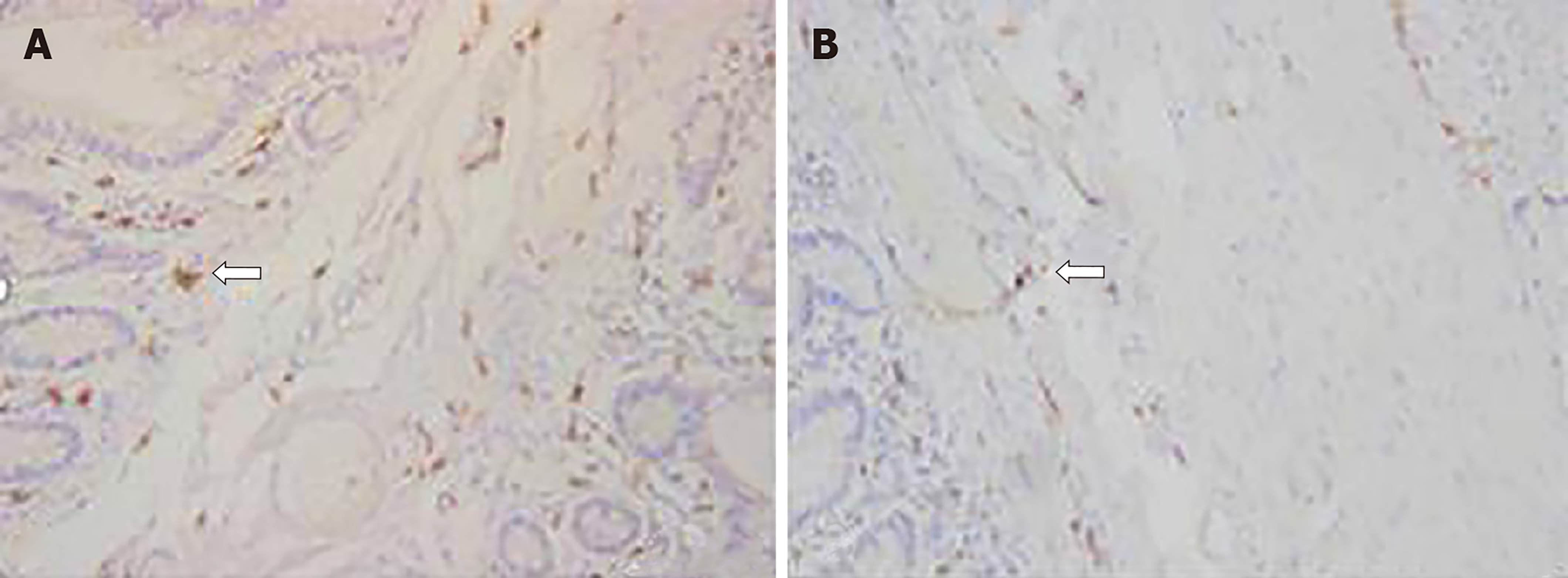
Figure 1 Immunohistochemical staining for c-Kit and Tenascin-X proteins.
A: High expression of c-Kit in the normal colon; B: Low expression of c-Kit in the slow transit constipation colon. The arrows indicate interstitial cells of Cajal showing brown-yellow c-Kit staining, suggesting that the number of interstitial cells of Cajal was reduced in the slow transit constipation group (200 ×).
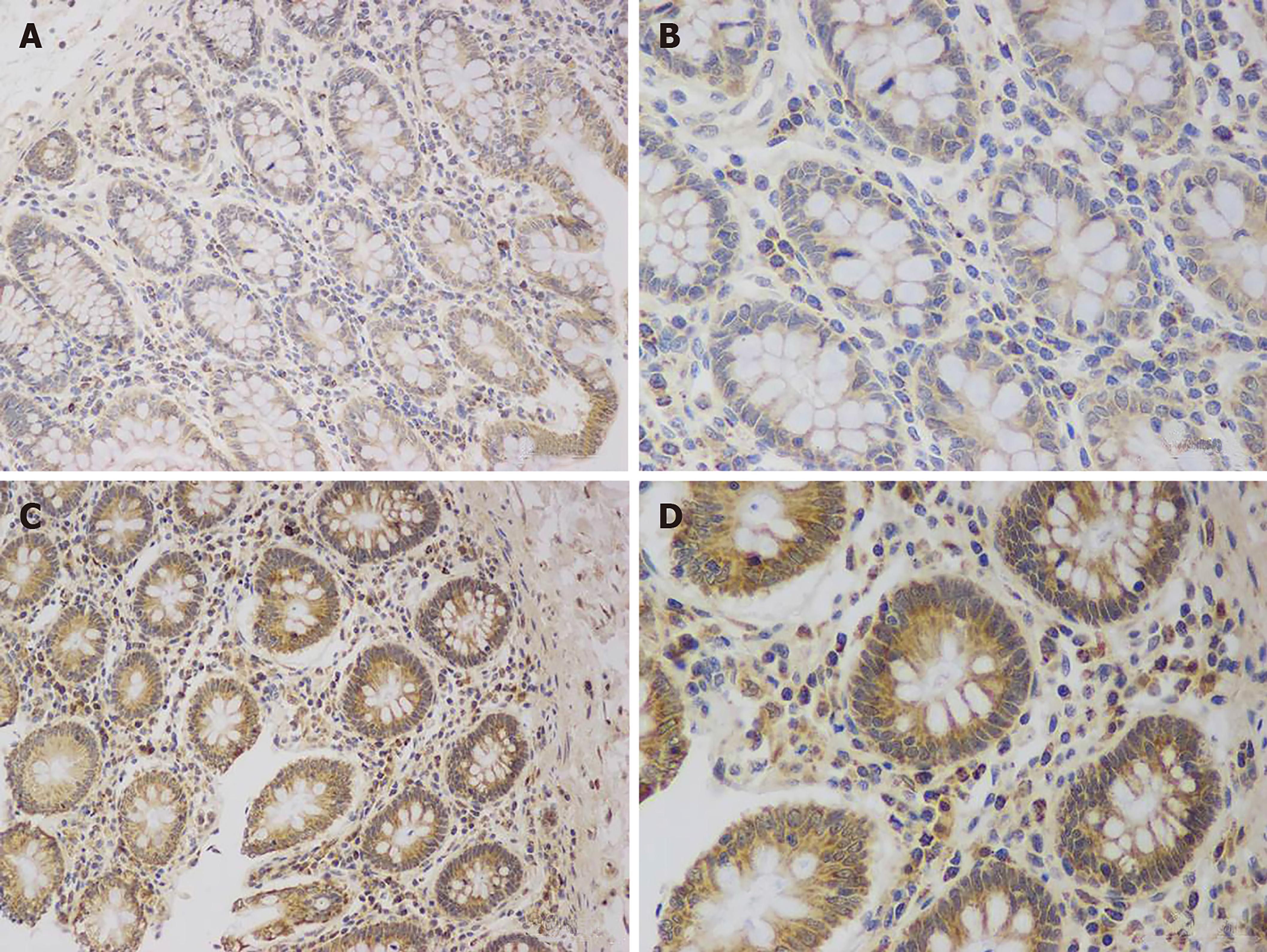
Figure 2 Immunohistochemical staining for c-Kit and Tenascin-X proteins.
A and B: Low expression of the Tenascin-X protein in the normal group (200 × and 400 ×, respectively); C and D: High expression of the Tenascin-X protein in the slow transit constipation group (200 × and 400 ×, respectively).
Figure 3 Expression of TNX, TGF-β, Smad2/3, and Smad7 in colon tissue.
The change in tenascin-X is consistent with changes in the TGF-β/Smad signaling pathway. NC: Normal group; STC: Slow-transit constipation group. aP < 0.01, bP < 0.001.
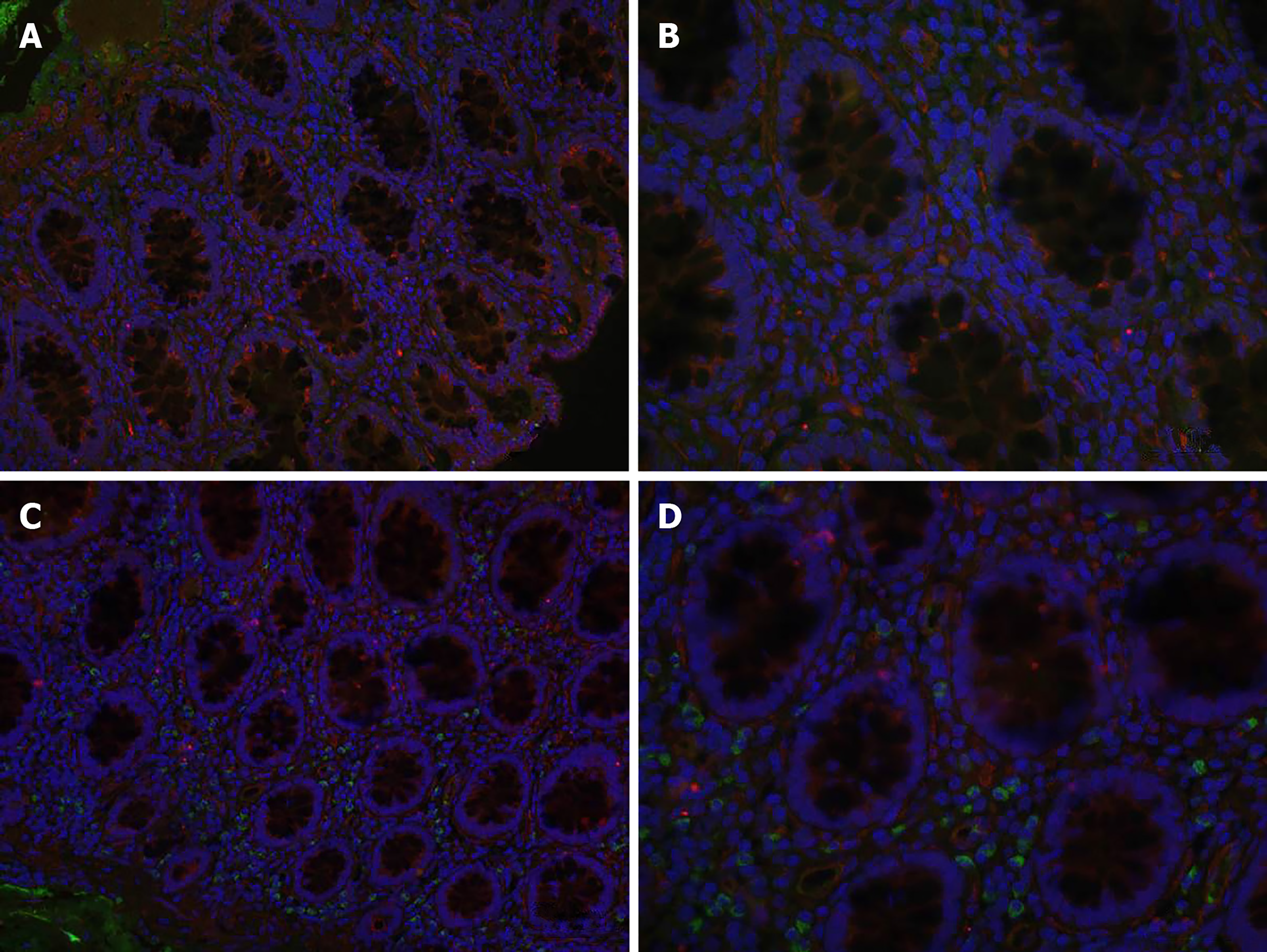
Figure 4 Expression of TGF-β and Smad2/3 in colon tissue.
A and B: Normal group (200 × and 400 ×, respectively); C and D: Slow transit constipation group (200 × and 400 ×, respectively).
- Citation: Zhang YC, Chen BX, Xie XY, Zhou Y, Qian Q, Jiang CQ. Role of Tenascin-X in regulating TGF-β/Smad signaling pathway in pathogenesis of slow transit constipation. World J Gastroenterol 2020; 26(7): 717-724
- URL: https://www.wjgnet.com/1007-9327/full/v26/i7/717.htm
- DOI: https://dx.doi.org/10.3748/wjg.v26.i7.717


