Copyright
©The Author(s) 2020.
World J Gastroenterol. Nov 28, 2020; 26(44): 7036-7045
Published online Nov 28, 2020. doi: 10.3748/wjg.v26.i44.7036
Published online Nov 28, 2020. doi: 10.3748/wjg.v26.i44.7036
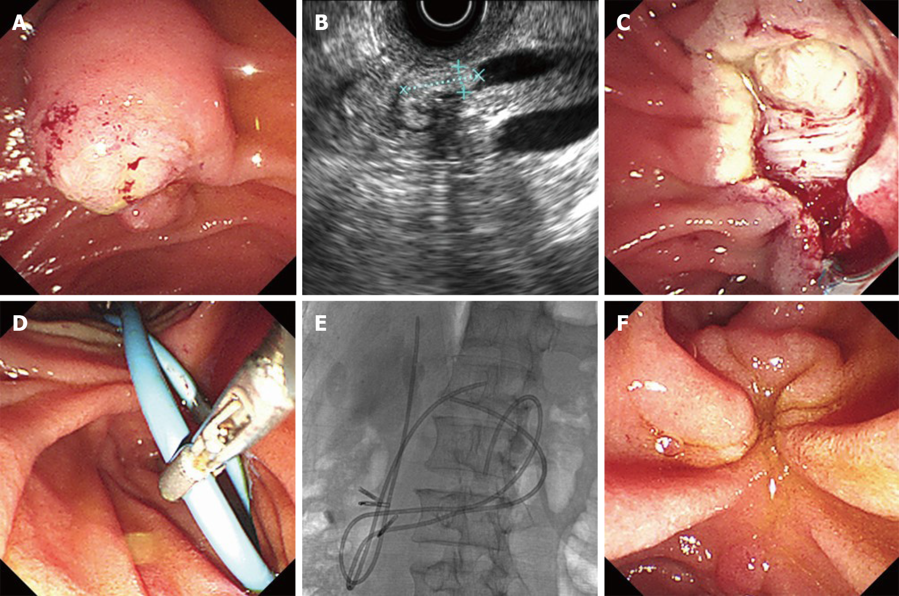
Figure 1 Endoscopic views of endoscopic papillectomy and overlength biliary and pancreatic stents placement in a patient with a lesion of the major duodenal papilla.
A: Duodenoscopy revealed an exposed-type tumor mass at the major duodenal papilla; B: Endoscopic ultrasonography showed an oval-shaped hyperechoic mass on the distal common bile duct; C: Injury of the muscularis propria was observed after endoscopic papillectomy; D: Overlength biliary and pancreatic stents were placed and adjusted; E: X-ray image showed overlength stents were successfully placed; F: A scar on the major duodenal papilla three months later.
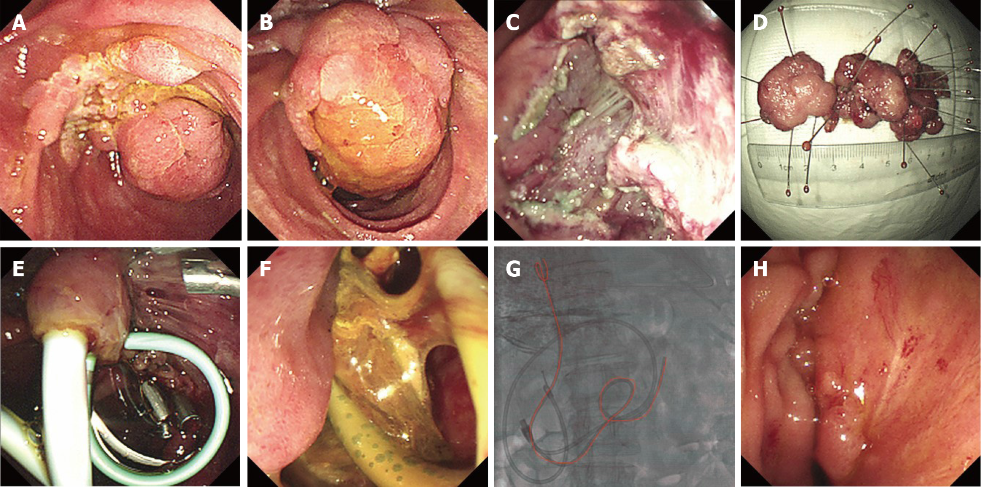
Figure 2 Overlength biliary stent in the treatment of delayed perforation after endoscopic papillectomy.
A: Duodenoscopy revealed a huge tumor mass at the major duodenal papilla (distant range view); B: Close range view of the huge tumor mass at the major duodenal papilla; C: Injury of the muscularis propria was observed after endoscopic papillectomy; D: View of the resected specimen, showing that it was 7 cm in length; E: Conventional biliary and pancreatic plastic stents were placed after endoscopic papillectomy; F: Delayed perforation and purulent necrosis were found; G: An overlength biliary stent was placed to drain bile to the proximal jejunum (shown in the red line); H: A scar on the major duodenal papilla three months later.
- Citation: Wu L, Liu F, Zhang N, Wang XP, Li W. Endoscopic pancreaticobiliary drainage with overlength stents to prevent delayed perforation after endoscopic papillectomy: A pilot study. World J Gastroenterol 2020; 26(44): 7036-7045
- URL: https://www.wjgnet.com/1007-9327/full/v26/i44/7036.htm
- DOI: https://dx.doi.org/10.3748/wjg.v26.i44.7036


