Copyright
©The Author(s) 2020.
World J Gastroenterol. Nov 28, 2020; 26(44): 6909-6922
Published online Nov 28, 2020. doi: 10.3748/wjg.v26.i44.6909
Published online Nov 28, 2020. doi: 10.3748/wjg.v26.i44.6909
Figure 1 The liver tissue is divided into nodules by fibrous septa of different widths.
Most of the hepatocytes in the nodules appear as bullous steatosis.
Figure 2 Edema of hepatocytes around the fibrous septum, a few of which showed balloon-like changes, and Mallory body can be seen.
A: Hematoxylin-eosin staining (HE), 200 ×; B: HE, 400 ×.
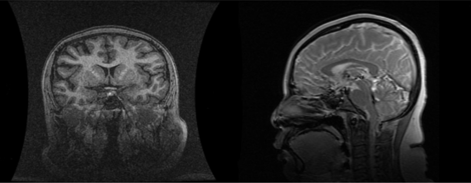
Figure 3 Cranial magnetic resonance: abnormal pituitary.
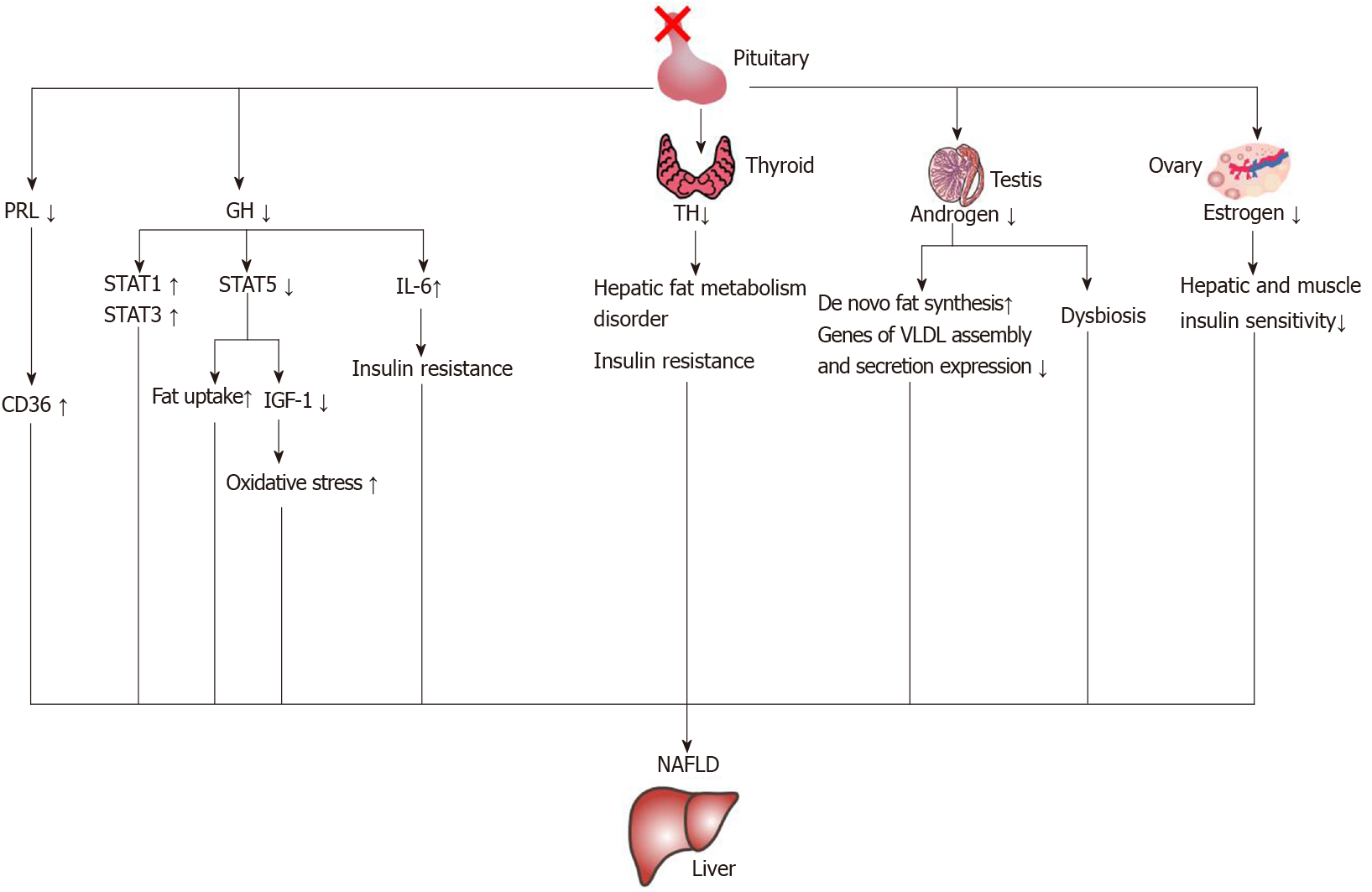
Figure 4 The mechanisms of nonalcoholic fatty liver disease induced by hormone deficiency.
IGF-1: Insulin-like growth factor-1; IL-6: Interleukin-6; NAFLD: Nonalcoholic fatty liver disease; PRL: Prolactin; GH: Growth hormone; STAT: Signal transducer and activator of transcription; TH: Thyroid hormone.
- Citation: Wu ZY, Li YL, Chang B. Pituitary stalk interruption syndrome and liver changes: From clinical features to mechanisms. World J Gastroenterol 2020; 26(44): 6909-6922
- URL: https://www.wjgnet.com/1007-9327/full/v26/i44/6909.htm
- DOI: https://dx.doi.org/10.3748/wjg.v26.i44.6909


