Copyright
©The Author(s) 2020.
World J Gastroenterol. Nov 14, 2020; 26(42): 6698-6705
Published online Nov 14, 2020. doi: 10.3748/wjg.v26.i42.6698
Published online Nov 14, 2020. doi: 10.3748/wjg.v26.i42.6698
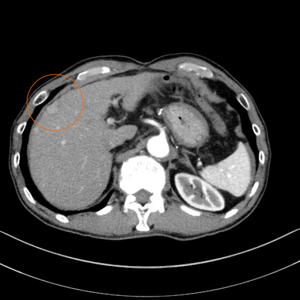
Figure 1 Abdominal contrast-enhanced computed tomography before the first surgery.
Arterial phase of abdominal contrast-enhanced computed tomography before the first surgery showed a tumor nodule 20 mm in diameter with early staining located in segment 8 of the liver (orange circle).
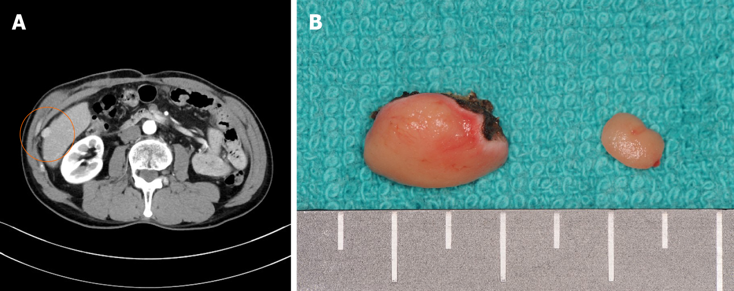
Figure 2 Abdominal contrast-enhanced computed tomography and the surgical specimen from the second surgery.
A: Arterial phase of abdominal contrast-enhanced computed tomography before the second surgery showed a tumor 10 mm in diameter, located in segment 6 (orange circle), and protruding to the surface of the liver with early staining; B: Surgical specimen of the liver tumor and peritoneal tumor at the second surgery.
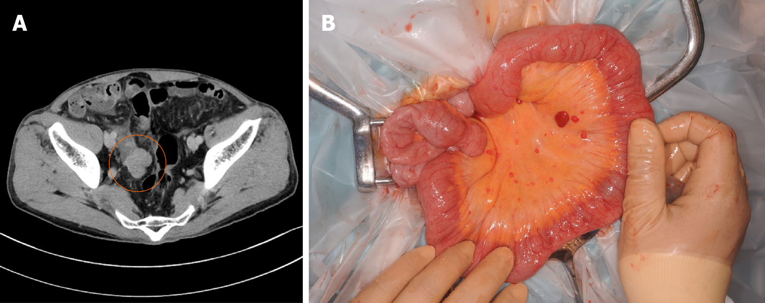
Figure 3 Abdominal contrast-enhanced computed tomography before the third surgery and the intraoperative findings.
A: Abdominal contrast-enhanced computed tomography showed a tumor 32 mm in diameter in the pelvis (orange circle); B: Many small peritoneal nodules were found at the time of laparotomy.
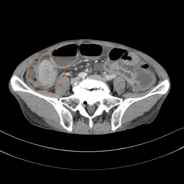
Figure 4 Abdominal contrast-enhanced computed tomography demonstrated an intussusception of the small intestine due to a well-defined, rounded, enhancing endoluminal mass (orange circle).
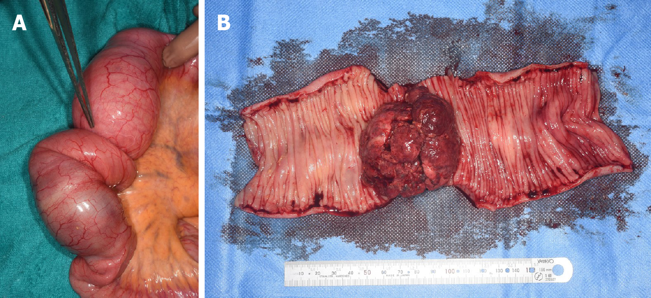
Figure 5 Intraoperative findings and the resected specimen.
A: The intussusception site was found 130 cm distal to the ligament of Treitz; B: The resected specimen showed a polypoid tumor 50 mm in diameter protruding into the lumen.
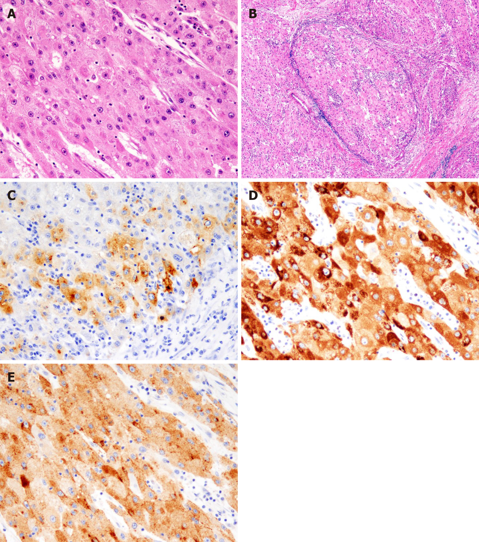
Figure 6 Histopathological findings and immunohistochemistry.
A: Histological findings showed tumor cells with cytoplasm rich in eosinophilic granules, enlarged nuclei, and clear nucleoli that showed dense proliferation on hematoxylin and eosin staining (× 400); B: Intravascular invasion of tumor cells were observed on Victoria blue staining (× 40); C: Alpha-fetoprotein (AFP) positive cells were observed on immunostaining (× 400); D: Hep Par1 positive cells were observed on immunostaining (× 400); E: Glypican3 positive cells were observed on immunostaining (× 400).
- Citation: Mashiko T, Masuoka Y, Nakano A, Tsuruya K, Hirose S, Hirabayashi K, Kagawa T, Nakagohri T. Intussusception due to hematogenous metastasis of hepatocellular carcinoma to the small intestine: A case report. World J Gastroenterol 2020; 26(42): 6698-6705
- URL: https://www.wjgnet.com/1007-9327/full/v26/i42/6698.htm
- DOI: https://dx.doi.org/10.3748/wjg.v26.i42.6698


